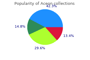
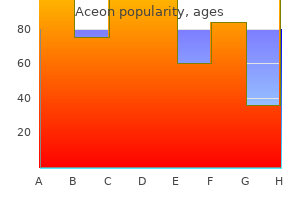
Aceon dosages: 8 mg, 4 mg, 2 mg
Aceon packs: 30 pills, 60 pills, 90 pills, 120 pills, 180 pills, 270 pills, 360 pills

Aceon 2 mg buy genericHowever hypertension knowledge test aceon 4 mg purchase online, life-threatening bleeding was more frequent with combination therapy than with antiplatelet remedy (4 blood pressure chart by race aceon 8 mg purchase otc. This inter-individual variation in warfarin dosing could reflect dif- 22 ferences in age blood pressure medication ok for pregnancy aceon 2 mg order on-line, weight heart attack back pain aceon 4 mg discount with visa, liver operate, food plan, alcohol consumption, concomitant medicines, and comorbid sicknesses. The decision to use warfarin alone or in combination with aspirin ought to be based mostly on a careful evaluate of the risks of future vascular and bleeding events, affected person compliance with therapy, and the provision of top quality warfarin monitoring. Although aspirin is prone to remain first-line therapy for most sufferers with coronary artery disease, warfarin therapy could also be helpful in higher risk patients and these who endure recurrent events regardless of aspirin remedy. Anticoagulants Side Effects Bleeding is the most frequent complication of warfarin therapy. The risk of bleeding is influenced by the depth of anticoagulation; the concomitant use of aspirin, nonsteroidal anti-inflammatory agents, or different medication that affect hemostasis; a history of bleeding; superior age; a historical past of stroke; or the presence of great comorbid circumstances. To circumvent this complication, sufferers with known protein C or protein S deficiency should be began on maintenance, somewhat than loading, doses of warfarin after therapeutic doses of heparin have been given. Because warfarin is teratogenic, its use should be avoided, if attainable, in being pregnant. Despite 248 promising knowledge, the function of the opposite agents on this affected person inhabitants remains to be clearly delineated. The biggest unmet want in anticoagulation therapy is replacement of warfarin with an orally lively agent that can be given in fixed doses with out routine coagulation monitoring. Consequently, a lot of the present consideration is concentrated on new oral anticoagulants. Those in essentially the most superior phases of improvement are the oral direct thrombin and issue Xa inhibitors. Dabigatran etexilate and rivaroxaban have been licensed for thromboprophylaxis in sufferers undergoing hip or knee substitute 22 surgery in Europe and Canada. The problem for the longer term might be to decide which of the numerous agents currently under improvement will provide the best efficacy with the greatest diploma of security. Fuster V: Elucidation of the position of plaque instability and rupture in acute coronary events. Yamamo to M, Nakagaki T, Kisiel W: Tissue factor-dependent autoactivation of human blood coagulation issue. Danielsson A, Raub E, Lindahl U, et al: Role of ternary complexes by which heparin binds both antithrombin and proteinase, within the acceleration of the reactions between antithrombin and thrombin or issue Xa. The International Study Group: In-hospital mortality and scientific course of 20,891 sufferers with suspected acute myocardial infarction randomised between alteplase and streptokinase with or with out heparin. Collins R, MacMahon S, Flather M, et al: Clinical effects of anticoagulant therapy in suspected acute myocardial infarction: Systematic overview of randomised trials. Holdright D, Patel D, Cunningham D, et al: Comparison of the effect of heparin and aspirin vs. Theroux P, Waters D, Lam J, et al: Reactivation of unstable angina after the discontinuation of heparin. Oldgren J, Grip L, Wallentin L: Reactivation after cessation of thrombin inhibition in unstable coronary artery disease, regardless of aspirin dose. Evidence for biological stabilization of issue Xa by factor V-phospholipid advanced. Basu D, Gallus A, Hirsh J, et al: A prospective research of the value of heparin therapy with the activated partial thromboplastin time. Antman E, Beasley J, Califf R, et al: American College of Cardiology/American Heart Association Task Force on Practice Guidelines. Amiral J, Bridey F, Wolf M, et al: Antibodies to macromolecular platelet factor 4-heparin complexes in heparin induced thrombocytopenia: A study of 44 circumstances. Young E, Cosmi B, Weitz J, Hirsh J: Comparison of the non-specific binding of unfractionated heparin and low-molecular-weight heparin (enoxaparin) to plasma proteins. Barzu T, Molho P, Tobelem G, et al: Binding and endocytosis of heparin by human endothelial cells in culture. Glick A, Kornowski R, Michowich Y, et al: Reduction of reinfarction and angina with use of low-molecular-weight heparin remedy after streptokinase (and heparin) in acute myocardial infarction. Comparison of lowmolecular-weight heparin with unfractionated heparin acutely and with placebo for eighty three. A comparability of lowmolecular-weight heparin with unfractionated heparin for unstable coronary artery illness. James S, Armstrong P, Califf R, et al: Safety and efficacy of abciximab mixed with dalteparin in acute coronary syndromes. Young J, Kereiakes D, Grines C, et al for the National Investigators Collaborating on Enoxaparin investigators: Low-molecular-weight heparin remedy in percutaneous interventions. Woltzt M, Weltermann A, Nieszpaur-Los M, et al: Studies on the neutralizing effects of protamine on unfractionated and low-molecular-weight heparin (Fragmin) at the web site of activation of the coagulation system in man. Monreal M, Lafoz E, Olive A, et al: Comparison of subcutaneous unfractionated heparin with low-molecular-weight heparin (Fragmin) in sufferers with venous thromboembolism and contraindications to Coumadin. Walenga J, Jeske W, Bara L, et al: Biochemical and pharmacological rationale for the event of an artificial heparin pentasaccharide. Paolucci F, Clavies M, Donat F, et al: Fondaparinux sodium mechanism of motion: Identification of specific binding to purified and human plasma-derived proteins. Matziolis G, Perka C, Disch A, et al: Effects of fondaparinux compared with dalteparin, enoxaparin and unfractionated heparin on human osteoblasts. Handschin A, Trentz O, Hoerstrup S, et al: Effect of low molecular weight heparin (dalteparin) and fondaparinux (Arixtra) on human osteoblasts in vitro. Mazzolai L, Hohfeld P, Spertini F, et al: Fondaparinux is a safe various in case of heparin intolerance during pregnancy. Fox I, Dawson A, Loyonds P, et al: Anticoagulant exercise of Hirulog, a direct thrombin inhibitor, in humans. Greinacher A, Lubenow N: Recombinant hirudin in scientific apply: Focus on lepirudin. Greinacher A, Volpel H, Janssens U, et al: Recombinant hirudin (lepirudin) offers secure and effective anticoagulation in sufferers with the immunologic kind of heparininduced thrombocytopenia: A prospective examine. Vanholder R, Dhondt A: Recombinant hirudin: Clinical pharmacology and potential functions in nephrology. Sorenson B, Ingerslev J: A direct thrombin inhibitor studied by dynamic complete blood clot formation. Lindhoff-Last E, Bauersachs R: Heparin-induced thrombocytopenia-alternative anticoagulation in pregnancy and lactation. Theroux P, Perez-Villa F, Waters D, et al: A randomized double-blind comparison of two doses of hirulog or heparin as adjunctive therapy to streptokinase to promote early patency of the infarct-related artery in acute myocardial infarction. Treatment with bivalirudin (hirulog) as in contrast with heparin throughout coronary angioplasty for unstable or post-infarction angina. Fitzgerald D, Murphy N: Argatroban: A artificial thrombin inhibitor of low relative molecular mass. Gustafsson D, Nystrom J-E, Carlsson S, et al: Pharmacodynamic properties of H376/95, a prodrug of the direct thrombin inhibitor melagatran, meant for oral use. Blech S, Ebner T, Ludwig-Schwellinger E, et al: the metabolism and disposition of the oral direct thrombin inhibitor, dabigatran, in humans. Connolly S, Ezekowitz M, Yusuf S, et al: Dabigatran versus warfarin in sufferers with atrial fibrillation. Abraham E, Reinhart K, Opal S, et al: Efficacy and safety of tifacogin (recombinant tissue factor pathway inhibitor) in severe sepsis: A randomized managed trial. Hinder M, Frick A, Jordaan P, et al: Direct and fast inhibition of issue Xa by otamixaban: A pharmacokinetic and pharmacodynamic investigation in sufferers with coronary artery disease. Late-breaking medical trial: a dose discovering examine of the oral direct issue Xa inhibitor apixaban in the treatment of patients with acute symptomatic deep vein thrombosis. The Persist Investigators: A novel long-acting synthetic factor Xa inhibitor (SanOrg34006) to replace warfarin for secondary prevention in deep vein thrombosis. The Van Gogh Investigators: Idraparinux versus commonplace therapy for venous thromboembolic disease. The Van Gogh Investigators: Extended prophylaxis of venous thromboembolism with idraparinux. Amadeus Investigators: Comparison of idraparinux with vitamin K antagonists for prevention of thromboembolism in patients with atrial fibrillation: A randomised, open-label, non-inferiority trial.
Buy discount aceon 8 mgThree imaging contrasts are used to decide plaque composition: (1) T1-weighted imaging; (2) T2-weighted imaging; and (3) proton density�weighted imaging prehypertension cure aceon 2 mg cheap without a prescription. Information obtained concerning the fee of vitality release (spin-lattice leisure time) and the rate at which the spins diphase (spinspin leisure time) are referred to as T1- and T2-weighted measurements arrhythmia heart murmur cheap aceon 8 mg on-line, respectively blood pressure medication used for hot flashes aceon 8 mg order without prescription. Furthermore arrhythmia strips effective aceon 4 mg, proton density� weighted imaging may be obtained by adjusting the imaging parameters to cut back T1 and T2 contributions, leaving solely the differences in water or lipid proton density. The use of in vitro standard magnetic resonance imaging techniques has been evaluated for the examine of atherosclerotic plaques in human carotid arteries,64,sixty five aorta,sixty six peripheral arteries,67 and coronary arteries. In contrast to typical magnetic resonance imaging of the carotid arteries, preliminary research in an animal pig mannequin have shortly demonstrated the constraints of assessing the coronary arteries by standard magnetic resonance imaging in vivo. The most necessary limitations included cardiac and respiratory motion artifacts and the deeply located and comparatively smaller and nonlinear course of the coronary arteries. To overcome the potential shortcomings of typical surface magnetic resonance imaging, placement of an intravascular receiver coil in combination with an external magnetic field is feasible. Intravascular coils have demonstrated good correlation between histologic sections and intravascular magnetic resonance imaging of human thoracic aortic segments. An essential safety concern is vessel heating and harm brought on by the vitality emitted by the intravascular coil. Although the coronary blood move might partially dissipate the warmth, smaller vessels may be more vulnerable to temperature will increase. Conventional magnetic resonance imaging, with or without intravascular coils, requires the application of exterior magnets and subsequently a magnetic suite. It has made such evaluations impractical in the cardiac catheterization laboratory. The threshold worth of temperature variation above which the rate of opposed cardiac occasions was significantly elevated was zero. Most of the cardiac occasions were related to restenosis of the handled lesion and to not a recurrent acute coronary syndrome. Thermography has permitted some fascinating observations on the nature of coronary plaques. In one small examine involving sufferers with stable and unstable coronary artery disease, roughly one third of the lesions had been found to be sizzling, indicating that plaque irritation was fairly prevalent. It was observed that sufferers with unstable coronary syndromes may have both cold and warm lesions solely millimeters apart in distinction to findings suggesting diffuse vessel inflammation in patients with unstable coronary syndromes. Some have instructed that correct interpretation of intravascular thermography knowledge requires further complementary information, such as coronary blood move and structural traits of the atherosclerotic plaques. At present, robust pathophysiologic knowledge support the function of heat manufacturing by weak plaques. The expertise to measure the warmth throughout the coronary plaques exactly is on the market. However promising this technology is, larger medical trials are required to decide the sensitivity and specificity earlier than it turns into broadly available. The depth of penetration of the field of view is roughly 50 to 200 �m in to the vessel wall. The probe accurately correlated with the histologic analysis in 15 of sixteen (94%) aortic lesions and in 16 of 18 (88%) coronary lesions. A first-in-man security and feasibility examine using the Topspin Medical probe has been accomplished in 29 sufferers. Six patients were excluded from evaluation due to poor-quality photographs because of artifacts. Molecular imaging of atherosclerosis coupled with magnetic resonance imaging may present alternative and extra promising modalities for imaging the vulnerable plaque. Although most of this scattered mild is on the identical wavelength because the incident mild, some is scattered at completely different wavelengths. This power distinction is called the Raman shift, defined by the 17 following formula: Emerging Diagnostic Procedures for the Vulnerable Plaque E v = E i - Es A plot of the incident light intensity versus Raman shift is a Raman spectrum. Different Raman spectrums are sometimes noticed, each related to the totally different vibrational or rotational motions of molecules in a pattern. When Raman spectroscopy is applied to an atherosclerotic plaque, the resultant spectrums can be thought-about to be a molecular fingerprint of that plaque. The identical group of investigators demonstrated the findings from Raman spectroscopy to correlate with ex vivo histologic specimens of atherosclerotic plaques obtained from human coronary and peripheral arteries. Furthermore, Raman spectroscopy detected reductions in cholesterol accumulation related to therapy with atorvastatin. A Raman spectroscopy catheter designed for the in vivo assessment of human coronary arteries has been developed but has not but been examined in human subjects. The catheter has an outer diameter of 2 mm and consists of a facet view�type micro-Raman spectroscopy probe, imaging fiber bundle, and balloon. Inflation of the balloon brings the probe closer to the lumen boundary for correct analysis. This limits picture acquisition to the fibrous cap and inside the atheromatous core. To date, experiments have been limited to direct contact with tissue; full noncontact circumferential imaging has not been evaluated. Spectroscopy is predicated on the principle that totally different chemical compounds take up and scatter totally different amounts of vitality at different wavelengths, leaving a singular chemical fingerprint. Currently, two forms of photonic spectroscopy present potential for the clinical detection of atherosclerotic and significantly susceptible plaques, Raman spectroscopy and near-infrared diffuse reflectance spectroscopy. Raman Spectroscopy Raman spectroscopy is a common analytic approach for identification of molecules in gases, liquids, and solids by the scattering of laser mild. Near-Infrared Diffuse Reflectance Spectroscopy Similar to Raman spectroscopy, near-infrared diffuse reflectance spectroscopy makes use of gentle to detect and decide the composition of natural substances. However, in distinction to Raman spectroscopy, which uses high-energy laser gentle within the visible gentle spectrum, near-infrared diffuse reflectance spectroscopy is the process of understanding how infrared light (750 to 2500 nm) interacts with various molecules. Near-infrared mild happens simply beyond the location of purple mild in the seen spectrum. The amount of sunshine absorbed is proportional to the focus of that particular molecule, revealing each qualitative and quantitative details about the pathologic process under investigation. Histologic correlation with reflectance patterns of various tissues can probably detect and distinguish the lipid-rich atheromatous core and turn into a useful diagnostic tool for the detection of susceptible plaque. Spectroscopic techniques utilizing infrared light have demonstrated the ability to determine ldl cholesterol, high-density lipoprotein, and low-density lipoprotein in arterial wall samples obtained at post-mortem. Chemical analyses have proven that correlation of the atheromatous core content using high-pressure liquid chromatography is excessive. Subsequently, the lipid content material of ex vivo specimens of human carotid plaques was successfully measured using near-infrared diffuse reflectance spectroscopy. These identical investigators found related sensitivities and specificities for the identification of lipid-rich plaques in a research of 167 human coronary artery specimens. In a research utilizing this device, large, lipid-rich plaques were identified (sensitivity, 88%; specificity, 79%) via up to three mm of blood. Although coronary artery movement might have an effect on the acquisition of knowledge by near-infrared diffuse reflectance spectroscopy, ultrafast systems have been developed which are able to obtaining spectra information within 6 milliseconds by scanning solely a preselected number of wavelengths applicable for atherosclerotic plaque assessment. Further technical advancements will address these issues and will enhance on the quality of knowledge and picture acquisition. Although they share the common goal of figuring out the weak plaque, they achieve this by concentrating on various parts of the atherosclerotic plaque. Future identification of a vulnerable plaque could result in better prognostic evaluations or therapy methods in sufferers with coronary artery illness. Yusuf S, Reddy S, Ounpuu S, Anand S: Global burden of cardiovascular diseases: Part I: General concerns, the epidemiologic transition, risk elements, and impact of urbanization. Naghavi M, Libby P, Falk E, et al: From vulnerable plaque to susceptible patient: Q name for brand spanking new definitions and risk evaluation strategies: Part I. Rioufol G, Finet G, Ginon I, et al: Multiple atherosclerotic plaque rupture in acute coronary syndrome: A three-vessel intravascular ultrasound study. Manfrini O, Mont E, Leone O, et al: Sources of error and interpretation of plaque morphology by optical coherence tomography. Nasu K, Tsuchikane E, Katoh O, et al: Accuracy of in vivo coronary plaque morphology assessment: A validation examine of in vivo virtual histology in contrast with in vitro histopathology.

Order aceon 2 mg free shippingYour candidacy is set on an individual foundation and includes many different elements arrhythmia uptodate cheap aceon 8 mg visa. After all of your exams have been accomplished blood pressure 120 0 purchase aceon 2 mg visa, your pretransplant heart specialist presents your case to a coronary heart transplant selection committee pulmonary hypertension 70 mmhg aceon 4 mg discount online. This is a staff that meets weekly and consists of heart surgeons arrhythmia symptoms in children aceon 4 mg buy low cost, cardiologists, transplant nurses, infection specialists, and a social worker. Your case is reviewed, and the committee discusses suggestions for the most effective course of therapy for you. In some circumstances you could be "too nicely" for itemizing right now and should continue on medicines. In other cases a patient could also be decided to not be a candidate who will benefit from a heart transplant, due to certainly one of several possible causes. Because there are so many more candidates for transplants than there are available hearts, the transplant listing have to be carefully screened. A cautious search is conducted for any potential contraindications to a heart transplant, such as undiagnosed most cancers or other serious medical problem. A description of most of the tests that are essential follows: Blood exams A sequence of blood exams are used to consider your liver function, kidney perform, blood and tissue type, and any earlier exposure to various infections. Small pads (electrode leads) related with wires are placed over completely different components of your body. These leads noninvasively detect the rhythm and sample of the electrical waves of your heart and convert it in to strains on a sheet of paper for your doctor to interpret. Echocardiogram (Echo) An echocardiogram is a noninvasive form of cardiac imaging (an ultrasound of the heart) that uses sound waves to study the scale, shape, and movement of all cardiac constructions. The important structures which would possibly be seen embrace the 4 heart valves, the pumping perform of the right and left ventricles (lower chambers), and the pericardial sac (the lining of the heart). This is finished to detect any lung illness and to determine your ability to wean from the respiration ventilator after your transplant surgical procedure. If the carotid arteries have severe narrowing, this have to be corrected before your transplant surgery. Abdominal ultrasound An ultrasound of your belly is a painless check performed to rule out any gallbladder disease/stones and to assess for an abnormally enlarged belly aorta (aneurysm). Lowenergy x-rays are handed through the bones to measure the mineral (calcium) content material of the bones. If wanted, a 24-hour urine assortment is completed to additional assess your kidney function. It is necessary to undergo a complete cancer screening as part of your transplant evaluation. Stool pattern to detect blood A simple stool sample check (a "stool guaiac card") is a simple approach to check the stool for any hidden blood within the intestinal tract. If necessary, a colonoscopy may be required to extra rigorously exclude any potential colon cancer. Left coronary heart catheterization and coronary angiogram that is described in additional detail in Question 74. A left heart catheterization permits your doctor to truly see how the blood flows by way of your heart and coronary arteries. It is often carried out through a small catheter (fine hollow tube) inserted in to an artery on the aspect of your groin (the femoral artery). This invasive check is performed to measure the pressures inside the proper aspect of your coronary heart and to check for the presence of pulmonary hypertension (elevated strain in the lungs). This is a particular kind of train test that measures your mixed heart and lung operate and ability to use oxygen. A coronary heart catheterization is an invasive heart test performed by a cardiologist using your arteries or veins to acquire details about your coronary heart and its perform. It may involve a left heart catheterization, a proper coronary heart catheterization, or both. Left Heart Catheterization and Coronary Angiogram A left heart catheterization allows your doctor to really see how the blood flows by way of your coronary heart and coronary arteries. This is the best way to evaluate the coronary arteries for any potential blockage problems. After rigorously cleaning and sterilizing the realm, a cardiologist inserts a catheter (a small, nice, hole tube) in to an artery on the side of your groin and uses an x-ray camera to guide the catheter as much as your coronary heart. You are awake but sedated during this routine process, which takes roughly 1 hour. After your transplant, additionally, you will have this procedure as part of your annual posttransplant check-up. A left heart catheterization is commonly accompanied by a proper heart catheterization (see below). It is done to examine for the presence of pulmonary hypertension (elevated pressure within the lungs). After fastidiously cleansing the facet of your neck, a heart specialist inserts a small catheter (a fine, hollow tube) in to the massive vein on the facet of your neck (the jugular vein). This special catheter has a soft inflatable balloon on the tip, generally recognized as a Swan-Ganz catheter or a pulmonary artery catheter. Some sufferers are so sick while ready for a heart transplant that they require a long-term pulmonary artery catheterization, whereby the Swan-Ganz catheter is left of their neck till they receive a brand new coronary heart. Their tenuous medical situation could require continual monitoring of their heart pressures to stop their clinical status from deteriorating whereas they await a new heart. Hemodynamics Pressures in every of your heart chambers and in your lungs (pulmonary artery pressure). This is named pulmonary hypertension and essentially is hypertension in the arteries that provide the lungs. The proper coronary heart catheterization (using a Swan-Ganz catheter) can precisely measure and record this strain. If your right heart catheterization demonstrates pulmonary hypertension, the cardiologist might need to think about additional testing throughout your catheterization. If the pulmonary pressures are too excessive, special intravenous medications (vasoactive drugs) are given to try to lower the pulmonary pressures or to enhance the forward pumping drive of the center (the cardiac output). They are referred to as vasoactive as a result of they sometimes work by performing on the blood vessels within the body, sometimes dilating (enlarging) them, which may act to decrease the pressure within the arteries. This might reduce the pulmonary artery stress and may enhance the cardiac output of the heart. These vasoactive medicines are given slowly in accordance with a standardized administration protocol. At every stage of the protocol, careful measurements of the hemodynamics are repeated and measured. Usually, the pulmonary pressures finally come down with escalating doses of the medicines, and the take a look at may then be concluded. If that is the case, these sufferers may not be eligible to obtain a coronary heart transplant. If pulmonary hypertension is current, then a vasoactive drug study might be carried out to show that it stays reversible. Cardiopulmonary train testing provides essential data in a patient who has coronary heart failure. Under regular circumstances a healthy body continues to improve oxygen intake and uptake because it increases its exercise intensity. Because of this established correlation in heart failure patients, your maximal oxygen consumption number is considered as part of your transplant analysis. Of course, as with your whole pretransplant testing, your case is considered individually, but that is still an necessary component of your evaluation. The syndrome of heart failure involves a reduced forward pumping of blood out of the heart (reduced cardiac output). A discount in enough blood flow to the kidneys can adversely have an result on how they perform. Additionally, some other medical circumstances that usually accompany coronary heart failure, such as high blood pressure and diabetes, can contribute and instantly influence the kidneys, which can additional compound the problem and might cause additional weakening of the kidney perform.
Aceon 4 mg cheap otcThe apical inferior phase may be supplied by the left anterior descending artery or the proper coronary artery blood pressure low diastolic 4 mg aceon purchase with mastercard. In the parasternal long-axis view hypertension facts purchase aceon 4 mg free shipping, the anterior interventricular septum is perfused by the left anterior descending artery prehypertension diet aceon 8 mg order online, the first 1 to 2 cm being perfused by the primary septal perforator arrhythmia frequency order aceon 4 mg on line, allowing dedication of whether or not the obstruction is proximal or distal to this left anterior descending department. In the parasternal short-axis view, the left anterior descending artery supplies the anterior wall and anterior septum, the circumflex artery provides the lateral wall, and the best coronary artery supplies the inferior septum and inferior wall. In the apical two-chamber view, the anterior wall is perfused by the left anterior descending artery, the inferior wall is supplied by the best coronary artery, and the apex usually has a twin coronary supply. In the apical four-chamber view, the midseptum is perfused by the left anterior descending artery, the basal septum is often a part of the best coronary artery territory, the apex is usually perfused by the left anterior descending artery, and the basal and midlateral partitions are equipped by the circumflex artery. A transmural infarction typically produces profound adjustments in regional left ventricular function, with most of the affected segments being akinetic or dyskinetic and the others being severely hypokinetic. In contrast, a nontransmural infarction leads to a lesser degree of hypokinesis and higher international ventricular operate. When the inferior wall myocardial infarction was complicated by a ventricular septal defect, echocardiography recognized involvement of the inferior portion of the interventricular septum despite the absence of electrocardiographic proof of septal involvement in these sufferers. One potential pitfall of transthoracic imaging is wrong positioning within the apical views, which truncates the true left ventricular apex, but the skilled echocardiographer ensures that the transducer is sufficiently low and lateral on the chest to avoid this problem. Echocardiographically, the aneurysmal segments are dyskinetic or akinetic and trigger distortion of the left ventricular form (with a wide neck), which persists in diastole. Almost 90% of true left ventricular aneurysms contain the apex, however extension to the anterior wall is frequent. This rare and doubtlessly life-threatening entity results from a rupture through the myocardium, with the extravasated blood being contained by the parietal pericardium. Pathologically, a small channel connects the left ventricle with a large blood- and thrombus-filled cavity lined by fibrous pericardial tissue, and a tear in the myocardium can be identified. Echocardiographically, an echo-free area outdoors the left ventricular cavity is seen related to it by a narrow neck, with an abrupt interruption within the ventricular wall. Rupture of the interventricular septum is more widespread with anterior than inferior infarcts. The perforation could additionally be a direct throughand-through hole or may be extra irregular and serpiginous Echocardiography After Reperfusion Therapy Multiple potential randomized trials have shown that the restoration of antegrade move after pharmacologic or mechanical reperfusion is normally related to improved wall motion, fewer problems, and decreased mortality. The extent of systolic function recovery is related to the duration of the occlusion (a prompt therapy begun in the first 2 hours after the onset of the chest pain offers the best results), extent of the ischemic zone, and success of reperfusion. Studies with sequential echocardiograms recommend, nevertheless, that restoration often occurs 24 hours to 10 days after reperfusion but may take 3 to 4 weeks if gorgeous is present. Echocardiography combined with dobutamine infusion (5 to 10 �g/kg/ min) can be used to distinguish surprised myocardium after thrombolytic therapy from nonviable myocardium, with the former responding to low-dose inotropic stimulation. Echocardiography often detects the septal defect directly as an interruption in the myocardium in an akinetic region, typically at the junction with regular or hyperkinetic tissue. Careful two-dimensional and color Doppler scanning of the ventricular septum is required in the course of the echocardiographic examination, particularly in the apical four-chamber and five-chamber views. Pulsed, continuous-wave, and colour Doppler affirm the left-to-right shunt throughout the septal defect. The defect size determined by shade Doppler echocardiography has been proven to correlate closely with that determined at surgical procedure or autopsy and with the pulmonic-to-systemic flow ratio measured at cardiac catheterization. Because its blood supply depends on a single coronary artery, the posteromedial papillary muscle is more regularly affected. Clinically, partial rupture of a papillary muscle head is seen more incessantly, as a result of complete rupture usually is rapidly deadly. The left ventricle is usually hyperdynamic in the presence of papillary muscle rupture, and this frequently renders the identification of a regional wall motion abnormality within the inferior wall troublesome. A, Apical two-chamber view shows rupture of the anterolateral papillary muscle, which moved freely in to the left atrium (arrow). An organized and nonmobile thrombus is detected within the first affected person (arrow in A), and a mobile thrombus (arrow in B) is seen in the second patient in a area of akinesia and apical aneurysm. The latter patient suffered a stroke inside hours of this echocardiographic examination. It could also be fixed, pedunculated, and freely cell, or might have a fixed base, with cell filaments extending from its surface. A newly formed ventricular thrombus is often solely mildly echogenic, but its appearance could additionally be speckled or contain areas brighter than the encompassing myocardium. The echogenicity can enhance, nonetheless, and calcification could additionally be discovered when the thrombus is organized. Because most left ventricular thrombi are within the apical region, normal and off-axis apical views (with optimized imaging of the area of interest) usually are required to verify their presence and distinguish them from a near-field artifact or a fibrous band (false tendon). When in contrast towards the more cumbersome but quantitative biplane Simpson technique, an tour of lower than 1. Many studies have compared echocardiographic methods with radionuclide ventriculography as a gold standard. The absence of such compensatory hyperkinesis suggests multivessel coronary illness and is associated with an increased incidence of dying, cardiogenic shock, development to a worse Killip class, and reinfarction. Similar results have been reported by Bolognese and colleagues111 and have been attributed to the no-reflow phenomenon. These results highlight the very fact 14 that the idea of successful reperfusion should include not solely early and sustained epicardial patency but in addition optimum tissue reperfusion. Myocardial contrast echocardiography is a promising methodology for the evaluation of myocardial blood move and could possibly be useful on this context. If the aim of exercise echocardiography is evaluation of regional wall movement only, treadmill train is often most well-liked. If additional Doppler information is desired, bicycle train allows assessment of each regional wall movement and Doppler during exercise, as a substitute of instantly after for the treadmill. Although vasodilators might have benefits for evaluation of myocardial perfusion, dobutamine is most popular for the assessment of regional wall movement. The enhance in systolic blood stress during the infusion may be more pronounced in hypertensive patients in comparison with normotensive patients. Patients with all three of these traits had a 13% probability of cardiac occasions within the first year and a higher threat throughout follow-up. The first is demonstration of myocardial viability in a hypokinetic or akinetic area, which suggests that regional systolic function will improve after revascularization in the case of hibernating myocardium or spontaneously on restoration from beautiful. The general sensitivity has ranged from 76% to 89% and the specificity from 70% to 95% for the detection of serious stenoses (50% narrowing of the arterial diameter) on coronary angiography. Vasodilator Stress Echocardiography Dipyridamole increases local adenosine ranges by inhibiting reuptake in to the endothelial cells. The mechanism of motion of dipyridamole as an agent to detect ischemia is believed to be a coronary steal, during which normal arteries respond by a maximal dilation, whereas arteries with significant stenosis have a decreased response, leading to circulate heterogeneity. Echocardiography may be much less sensitive to mild levels of ischemia however is extra particular. Although pharmacologic stress echocardiography could seem to be a extra elegant approach, treadmill train echocardiography supplies necessary medical data by combining evaluation of power expenditure with the imaging technique. Advantages of stress echocardiography compared with other methods embrace shorter imaging time, lack of ionizing radiation, portability, immediate availability of the results, decrease value, and availability of other details about chamber size and function, wall thickness, valvular operate, pericardial effusion, and aortic root disease. Tardif and colleagues have proven that the left primary coronary artery with its bifurcation might be visualized in all patients with a sensitivity of one hundred pc for detection of coronary narrowing in contrast with angiography. The stenosis severity 143 could also be underestimated with angiography because the reference section with which the stenosis is compared may be concerned in the diffuse atherosclerotic course of. Echocardiography in Acute Coronary Syndromes visualized in 69% and 31%, the proximal and midsegments of the circumflex artery in 80% and 51%, and the corresponding segments of the best coronary artery in 84% and 16% of sufferers. Anderson and coworkers181 have proven that endothelial function measured at the brachial artery stage correlates carefully with that in the coronary arteries. There is mild angiographic luminal narrowing (distal arrow) when the lesion is compared with a pseudonormal reference segment (proximal arrow). On intravascular ultrasound, the reference phase reveals significant plaque burden (central panel), and the severity of the goal lesion (right panel) is best appreciated. Theroux P, Ross J Jr, Franklin D, et al: Regional myocardial function within the acutely aware dog throughout acute coronary occlusion and responses to morphine, propranolol, nitroglycerin, and lidocaine. Zabalgoitia M, Ismaeil M: Diagnostic and prognostic use of stress echo in acute coronary syndromes together with emergency division imaging. Berning J, Steensgaard-Hansen F: Early estimation of risk by echocardiographic willpower of wall motion index in an unselected population with acute myocardial infarction.
Diseases - Short stature dysmorphic face pelvic scapula dysplasia
- Poikilodermatomyositis mental retardation
- Al Gazali Hirschsprung syndrome
- Gelineau disease
- Catel Manzke syndrome
- Renoanogenital syndrome
- Epilepsy, partial, familial
- Hereditary myopathy with intranuclear filamentous

2 mg aceon mastercardMasuyama T blood pressure chart by age nhs aceon 2 mg with visa, Kodama K blood pressure treatment guidelines aceon 4 mg discount line, Nakatani S blood pressure normal or high aceon 8 mg with amex, et al: Effects of adjustments in coronary stenosis on left ventricular diastolic filling assessed with pulsed Doppler echocardiography pulse pressure fitness cheap 8 mg aceon with visa. Norell M, Lythall D, Coghlan G, et al: Limited worth of the resting electrocardiogram in assessing sufferers with current onset chest ache: Lessons from a chest pain clinic. Task Force on Echocardiography in Emergency Medicine of the American Society of Echocardiography and the Echocardiography and Technology and Practice Executive Committees of the American College of Cardiology. Sasaki H, Charuzi Y, Beeder C, et al: Utility of echocardiography for the early evaluation of patients with nondiagnostic chest ache. Romano S, Dagianti A, Penco M, et al: Usefulness of echocardiography within the prognostic evaluation of non-Q-wave myocardial infarction. Relationship between systolic wall thinning and regional myocardial perfusion in extreme coronary stenosis. Porter A, Strasberg B, Vaturi M, et al: Correlation between electrocardiographic subtypes of anterior myocardial infarction and regional abnormalities of wall movement. Errichetti A, Homma S, Guyer D: Limitations of the 12-lead electrocardiogram in predicting segmental apical dysfunction: Comparison with apical dyfunction by 2-D echocardiography. Spiri to P, Bellotti P, Chiarella F, et al: Prognostic significance and pure historical past of left ventricular thrombi in sufferers with acute anterior myocardial infarction: A twodimensional echocardiographic study. Chockalingam A, Gnanavelu G, Alagesan R, Subramaniam T: Myocardial performance index in evaluation of acute right ventricular myocardial infarction. Sevimli S, Gundogdu F, Aksakal E, et al: Right ventricular pressure and pressure price properties in sufferers with proper ventricular myocardial infarction. Domingo E, Alvarez A, Garcia del Castillo H, et al: Prognostic worth of segmental contractility assessed by cross-sectional echocardiography in first acute myocardial infarction. Bigi R, Desideri A, Galati A, et al: Incremental prognostic worth of stress echocardiography as an adjunct to train electrocardiography after uncomplicated myocardial infarction. Heupler S, Mehta R, Lobo A, et al: Prognostic implications of train echocardiography in girls with recognized or suspected coronary artery illness. Previtali M, Fetiveau R, Lanzarini L, et al: Prognostic worth of myocardial viability and ischemia detected by dobutamine stress echocardiography early after acute myocardial infarction handled with thrombolysis. Perrone-Filardi P, Pace L, Prastaro M, et al: Assessment of myocardial viability in patients with continual coronary artery illness. Sitges M, Pare C, Azqueta M, et al: Feasibility and prognostic worth of dobutamineatropine stress echocardiography early in unstable angina. Weidemann F, Dommke C, Bijnens B, et al: Defining the transmurality of a continual myocardial infarction by ultrasonic strain-rate imaging: implications for figuring out intramural viability: An experimental study. Bjork Ingul C, Rozis E, et al: Incremental worth of pressure price imaging to wall motion evaluation for prediction of end result in patients present process dobutamine stress echocardiography. Hoffmann R, Altiok E, Nowak B, et al: Strain fee measurement by Doppler echocardiography allows improved assessment of myocardial viability inpatients with depressed left ventricular perform. Hoffmann R, Altiok E, Nowak B, et al: Strain rate evaluation allows detection of differences in diastolic function between viable and nonviable myocardial segments. Hanekom L, Jenkins C, Jeffries L, et al: Incremental worth of pressure rate analysis as an adjunct to wall-motion scoring for assessment of myocardial viability by dobutamine echocardiography: A follow-up research after revascularization. Vitarelli A, Montesano T, Gaudio C, et al: Strain fee dobutamine echocardiography for prediction of restoration after revascularization in patients with ischemic left ventricular dysfunction. Bolognese L, Sarasso G, Aralda D, et al: High-dose dipyridamole echocardiography early after uncomplicated acute myocardial infarction: Correlation with train testing and coronary angiography. Bolognese L, Rossi L, Sarasso G, et al: Silent versus symptomatic dipyridamoleinduced ischemia after myocardial infarction: Clinical and prognostic significance. Camerieri A, Picano E, Landi P, et al: Prognostic worth of dipyridamole echocardiography early after myocardial infarction in aged sufferers. Ismaeil M, Trusevich T, Nottestand S: Dobutamine esophageal echo in the assessment of coronary artery disease: Comparison with dobutamine transthoracic echo in the identical setting. Matsumura Y, Hozumi T, Arai K, et al: Non-invasive assessment of myocardial ischaemia using new real-time three-dimensional dobutamine stress echocardiography: Comparison with conventional two-dimensional methods. Aggeli C, Giannopoulos G, Misovoulos P, et al: Real-time three-dimensional dobutamine stress echocardiography for coronary artery disease analysis: Validation with coronary angiography. Peteiro J, Pinon P, Perez R, et al: Comparison of 2- and 3-dimensional exercise echocardiography for the detection of coronary artery illness. A new method for submit processing to 3-/4-dimensional images from three standard apical planes. Koenig K, Kasper W, Hofmann T, et al: Transesophageal echocardiography for diagnosis of rupture of the ventricular septum or left ventricular papillary muscle throughout acute myocardial infarction. Adachi H, Kyo S, Takamo to S, et al: Early diagnosis and surgical intervention of acute aortic dissection by transesophageal shade flow mapping. Yoshida K, Yoshikawa J, Hozumi T, et al: Detection of left primary coronary artery stenosis by transesophageal color Doppler and two-dimensional echocardiography. Intravascular ultrasound imaging in peripheral arterial and coronary artery illness. Physicians also increasingly are requested to use medical resources in a means that minimizes the financial impact. Conversely, low-risk sufferers are unlikely to profit from any intervention, especially one that itself has dangers of complications. These components represent two interactive influences, the extent of permanent damage and the extent of future myocardium in danger. Cardiac mortality elevated, especially after the ejection fraction decreased to lower than 40%. The variations had been biggest at the low end (ejection fraction < 20%), possibly reflecting the ameliorative results of angiotensin-converting enzyme inhibitor and beta blocker remedy that grew to become the usual of care in the Nineteen Nineties. Several other research have confirmed the necessary prognostic worth of ejection fraction in sufferers receiving thrombolysis. Simoons and colleagues4 discovered that 5-year survival was solely roughly 40% in patients with left ventricular ejection fraction lower than 30% in contrast with greater than 90% survival when the ejection fraction was larger than 40%. Event-free survival was approximately 75% for sufferers with an ejection fraction of 40% in contrast with survival lower than 25% for sufferers with an ejection fraction less than 40%. This suggests that the rise in ejection fraction at 6 weeks displays a decision of myocardial gorgeous. Conversely, 19% of sufferers showed a decrease in ejection fraction of 8% by 6 weeks. It is obvious that ejection fraction, reflecting the extent of scar, is a powerful predictor of mortality, but it displays what has already occurred. There is an important interplay between the extent of permanent injury (scar) and that of extra viable myocardium at risk. Ladenheim and associates12 found that among scientific and scintigraphic indices, the variety of reversible perfusion defects on stress thallium-201 photographs was one of the best predictor of future cardiac occasions. Compared with scientific or coronary angiography, these indices have been significantly more sensitive for detecting patients in danger for cardiac events. In-hospital cardiac occasions occurred in 43% of sufferers with vital reversible defects compared with 9% without vital reversibility. Over a mean 16-month follow-up, one of the best predictors of cardiac events had been extent of reversible 201Tl defects and ejection fraction. This alternative is particularly helpful for patients with contraindications to vasodilators, such as bronchospasm, current caffeine or methylxanthine exposure, high-degree atrioventricular block, or hypotension. In distinction to vasodilators, dobutamine produces a method more marked enhance in heart fee and blood pressure, leading to induction of true ischemia within the setting of coronary lesions, somewhat than simply heterogeneity of hyperemia. In this small choose cohort of seventy five patients, there have been no serious dobutamine-related issues. In contrast to train, vasodilator stress induces solely modest changes in determinants of myocardial oxygen demand. In addition, figuring out high-risk patients and directing appropriate therapy sooner can forestall early cardiac occasions. Nine of 20 patients (45%) with infarct zone reversible defects had in-hospital ischemic cardiac occasions in contrast with zero of 30 patients without (P =. Over a mean 12-month follow-up period, there have been three extra cardiac occasions in patients with reversible defects, whereas patients with out reversible defects remained freed from cardiac occasions. This was manifested as a greater ability to separate low-risk from high-risk sufferers.
Discount aceon 2 mg on-lineThe mostly used proton pump inhibitors are omeprazole (Prilosec) heart attack and blood pressure aceon 4 mg generic line, lansoprazole (Prevacid) pulse pressure 26 aceon 2 mg purchase overnight delivery, esomeprazole (Nexium) arrhythmia triggers aceon 4 mg buy otc, pantoprazole (Protonix) arteria brachialis purchase 4 mg aceon fast delivery, and rabeprazole (Aciphex). They are very well tolerated and extremely effective in treating ulcers and heartburn. High Blood Pressure Medications (Antihypertensives) People who take hypertension medications earlier than surgery are more probably to continue to need these medications to lower their blood pressure after surgical procedure. In addition, some people who had regular blood stress before surgical procedure could have hypertension after a transplant. Both cyclosporine and tacrolimus cause hypertension in about 70% of folks that take them. The mostly prescribed medicines for hypertension include diltiazem (Cardizem, Cartia, Dilacor, Tiazac), enalapril (Vasotec), lisinopril (Zestril), nifedipine (Procardia, Adalat), atenolol (Tenormin), and metoprolol (Lopressor). Notes About Antihypertensive Medications You may be suggested to follow a low-sodium food plan when you have or develop high blood pressure. Check with your transplant team earlier than starting or stopping any hypertension treatment. Possible Side Effects Dizziness and lightheadedness for the first few days, fatigue, nausea, loss of urge for food, headache, rash, dry cough, swelling within the ft, sluggish pulse, excessive potassium levels, kidney dysfunction. Low Blood Pressure Medications If your blood pressure is simply too low, your doctor may prescribe medicine to increase it. The medication mostly prescribed for low blood strain is fludrocortisone (Florinef). Notes About Fludrocortisone Fludrocortisone raises blood pressure by serving to you to retain salt in your physique and to discard potassium in your urine. Possible Side Effects Swelling in the palms or feet, fast weight achieve, water retention, headache. Spironolactone (Aldactone) might have been certainly one of your diuretics before transplantation-this medicine should be used very cautiously after transplantation. Potassium dietary supplements may be prescribed for a quick time to replenish the availability in your blood. Possible Side Effects Low blood strain, dizziness, lightheadedness, dehydration, more frequent urination, low potassium. Cholesterol-Lowering Medications Lowering your ldl cholesterol could assist prevent heart illness. The mostly used cholesterol-lowering medicines are atorvastatin (Lipitor), simvastatin (Zocor), rosuvastatin (Crestor), pravastatin (Pravachol), and lovastatin (Mevacor). The major immunosuppressive brokers, cyclosporine and sirolimus, are the culprits causing high ldl cholesterol and triglyceride ranges in many sufferers who take them. Notes About Cholesterol-Lowering Medications Cholesterol-lowering medicines often are taken at night. Possible Side Effects Upset stomach, heartburn, change in the greatest way meals style, diarrhea, skin rash, headache, constipation, blurred vision, muscle damage. Drug Interactions Some medicines can intervene with the greatest way cyclosporine, sirolimus, and tacrolimus are processed in your physique and can lead to very high or very low blood levels of those medication. Be positive to talk about possible drug interactions with any physician who prescribes a new drugs for you. In doing so, they might additionally tell your immune system to accept other foreign invaders that it ordinarily would battle. As a consequence, taking antirejection medications can place you at higher danger for developing an an infection. The commonest infections result from viruses that have been lying dormant in your system or within the donated organ. If an an infection is suspected, your caregivers might take sputum (the substance coughed up from your lungs), blood, and urine samples in addition to samples out of your catheter, wound, and drain sites. Signs that you may discover embrace fever, tiredness or fatigue, diarrhea or vomiting, redness or drainage around your incision or tube site, or a cough and sore throat. The infectious illness specialist works with the transplant group to manage and deal with infections. However, some people have to be readmitted to the hospital for therapy with intravenous drugs. The targets of the immune response are completely different from the targets in acute rejection. This damage happens very slowly and Chronic rejection Occurs a minimal of three months after transplant. Chronic rejection has options on tissue biopsy that are distinct from acute rejection, drug toxicity and different illnesses. Thanks to earlier recognition of acute and persistent rejection and the introduction of more powerful immunosuppressive agents (for instance, tacrolimus and sirolimus), many of those instances may be efficiently reversed. This longer survival comes at a worth, however: far more long-term issues than have been seen in the past. More attention must due to this fact be paid to the long-term effects of the immunosuppressive medicine and their cumulative effects. Over the course of a few years this mixture results in coronary heart assault and stroke. When expected survival after transplantation was quick, these long-term issues were of minimal concern. Today, with longer survival being commonplace, coronary heart disease is certainly one of the major causes of dying in transplant recipients. The incidence of hypertension is attributed to the first immunosuppressive agents. Standard antihypertensive medicines are efficient in treating this complication. Because many sufferers suffer from malnutrition before transplantation, these people are counseled to improve their nutrition afterward to help the healing process. Unfortunately, patients may turn out to be accustomed to this elevated calorie consumption and have a tough time chopping back their meals consumption as quickly as recovery from surgery has been achieved. The subsequent weight problems can lower mobility and enhance the chance of coronary artery disease. Once again, the offender is commonly the immunosuppressive agents, significantly tacrolimus and prednisone. The incidence of weight problems correlates with diabetes incidence and both are cardiac threat elements. Transplant-associated lymphoma is a feared complication of the immunosuppressive medicine. Chronic renal failure is a acknowledged complication of all organ transplantation because of the necessity for immunosuppression. Both tacrolimus and cyclosporine can cause the kidneys to function lower than optimally. Additionally, in patients with cirrhosis, heart disease, or renal disease earlier than transplantation, the unwanted effects of diuretic use, hypertension, and diabetes can all contribute to chronic renal failure in recipients of a new organ. Renal failure after the transplantation complicates medical management, resulting in increased morbidity and mortality. The incidence of continual renal illness amongst recipients of liver transplants is roughly 8% after 1 yr, 14% after three years, and 18% after 5 years. Some sufferers do, certainly, progress to dialysis; kidney transplantation could also be indicated in these people. A number of elements could predict the danger of developing renal failure, together with age (older patients have a higher risk), gender (males have a higher danger than females), pretransplantation kidney perform, and presence or absence of pretransplantation hypertension, diabetes, or hepatitis C an infection. Overall, non-White, non-African American sufferers have the bottom danger of continual renal failure. One approach is to scale back the dose of the primary immunosuppressive agent (that is, tacrolimus or cyclosporine). For these sufferers with a high threat or history of rejection, mycophenolate (CellCept, Myfortic) can be added to the drug routine. A current addition to the immunosuppression armamentarium, sirolimus (Rapamycin, Rapamune), can also reduce the danger.
Aceon 2 mg genericPlasma clearance of tirofiban is 20% to 25% decrease in older sufferers (65 years old) and can be decreased by 50% or more in patients with marked renal insufficiency (creati nine clearance <30 mL/minute) blood pressure medication for ptsd aceon 4 mg purchase line. Three doses of tirofiban have been studied in part I clinical trials: bolus dose of 5 blood pressure medication for diabetics aceon 2 mg buy cheap, 10 blood pressure video 4 mg aceon buy mastercard, or 15 �g/kg followed by a continu ous intravenous infusion of 0 pulse pressure 31 order aceon 8 mg without a prescription. A dosedependent inhibition of ex vivo platelet aggregation was observed within several minutes of bolus admini stration with sustained inhibition in the course of the maintenance infusion. Plasma clearance of tirofiban is decreased substantially in sufferers with extreme renal impairment (creatinine clearance <30 mL/minute), including sufferers requiring hemodialysis. The pharmacokinetics of eptifibatide are linear, and plasma concentrations are proportional to dose after intrave nous administration of 90 to 250 �g/kg and infusions of 0. Plasma concentrations of eptifibatide decline in a biexpo nential method after intravenous administration. Clear ance of eptifibatide is proportional to body weight and creati 9 clearance and inversely proportional to age. A double bolus technique (180 �g/kg, administered twice, 10 minutes apart) achieved maximal inhibition in a larger proportion of sufferers. Appropriate dosing of eptifibatide is based on creatinine clearance, a extra correct estimate of renal operate than serum creatinine alone. Patients with a creatinine clearance of <50 mL/min should receive an infusion of 1 �g/kg/min representing a 50% reduction of the normal infusion. The early reduc tion in mortality with aspirin continued when the patients had been noticed for a mean of 15 months. Aspirin reduced the risk of nonfatal reinfarction by 49% and nonfatal stroke by 46%. In particular, amongst patients younger than 70 years of age, the mixture markedly reduced mortality from 23. Overall, when all transfused, fatal, or cerebral bleeds had been 20 thought of collectively, there was no important excess risk asso ciated with using clopidogrel (134 [0. Antiplatelet remedy was associated with a extremely vital 15% relative discount in vascular deaths (P =. Overall, the relative odds of experiencing a significant extracranial hemorrhage was elevated 60% with antiplatelet therapy (odds ratio, 1. The enhance in deadly hemorrhage was not considerably totally different from that for nonfatal hemorrhage, though solely the surplus of nonfatal hemorrhagic occasions achieved statistical significance. The optimum dose of aspirin for the prevention of cardio vascular occasions has not been definitively established by directly comparing two different dosages in large medical trials. The up to date metaanalysis does, nonetheless, present useful information on the effects of various doses of aspirin. Overall, among 3570 sufferers in three trials instantly compar ing aspirin doses (75 mg vs. Considering each direct and oblique compari sons of aspirin dose, vascular events have been decreased 19% with 500 to 1500 mg/day, 26% with 160 to 325 mg/day, and 32% with seventy five to a hundred and fifty mg/day. These data present indirect support 210 for administration of an aspirin dose of seventy five to one hundred mg/day for heart problems therapy. Major bleeding (defined as disabling hemorrhage, intra ocular hemorrhage resulting in visible loss, or bleeding requir ing transfusion of at least 2 items of blood) was considerably more common in clopidogreltreated sufferers (3. There was not an excess price of deadly bleeding, bleeding that required surgical intervention, or hemorrhagic stroke. The variety of patients requiring trans fusion of two models of blood was greater in the clopidogrel group (2. The fee of major bleeding with clopidogrel was greater early (within 30 days of randomization; 2. Overall, the chance of minor bleeding was considerably larger in patients handled with clopidogrel (5. The two most just lately developed and studied P2Y12 receptor antagonists, prasugrel and ticagrelor shall be dis cussed later within the section "Emerging PlateletDirected Pharmacotherapy. Severe bleeding and transfusions were increased by approximatedly 40% with highdose clopidogrel compared to normal dosing. The general profit past 30 days is unknown (European Society of Cardiology, Barcelon, Spain, 2009). From day 29 via 12 months, sufferers within the loadingdose group obtained clopidogrel (75 mg/day), while these within the control group acquired placebo. Longterm aspirin therapy is recommended for Platelet-Directed Therapy Following Percutaneous CoronaryIntervention. An American Heart Association Science Advisory careworn the importance of no much less than 12 months of uninterrupted twin antiplatelet therapy and the schooling of sufferers and provid ers about the potential hazards related to premature discontinuation of those medication. In contrast, when stratified by hemorrhagic risk that included age sixty five years, prior stroke, history of bleeding, hema tocrit of less than 30%, diabetes mellitus, and a serum creati 9 larger than 1. Use of single antiplatelet therapy was more widespread in Europe than in the United States (34% vs. Similarly, the bottom efficient aspirin dose ought to be employed with combination therapies. Clinicians ought to consider proton pump inhibitors, significantly among sufferers with danger factors or a prior history of gastritis. Early eptifi batide administration was associated with larger charges of bleeding and pink cell transfusion. Emerging Platelet-Directed Pharmacotherapy the event of plateletdirected pharmacotherapy with optimized properties to include stronger platelet inhibi tion, in addition to rapidonset and rapidoffset pharmacodynam ics is proceeding rapidly. One or more brokers are poised to enter the world of patient care in the close to future. A post hoc analyses revealed either no benefit or potential harm from prasugrel in contrast with clopidogrel therapy in three affected person subgroups: patients older than seventy five years of age (no net scientific benefit); sufferers lower than 60 kg in body weight (no web medical benefit); and patients with a historical past of cerebrovascular events (prior ischemic stroke or transient ischemic attack) (net harm). The ratio is the chances ratio, quite than the hazard ratio, and was evaluated with using the Cochran-Mantel-Haenszel check. While largescale advanced biomarker and pharmacogenetic research are more doubtless to higher characterize and determine patients at risk for hemor rhagic issues, recurring ischemic/thrombotic occasions, drug failures and offtarget toxicities, platelet efficiency measurement instruments, if available to practicing clini cians and capable of providing information that might be translated directly to remedy determination, would also impression care meaningfully (reviewed in reference 96). For example, although aspirin reduces the danger of thrombotic occasions in highrisk patients by about 25%, 10% to 20% of treated sufferers may have another thrombotic event throughout longterm followup. These patients might require extra plateletdirected therapy with clopidogrel or different brokers. A key query referring to this issue is whether standardized laboratory exams assessing the platelet response to aspirin or clopidogrel can predict clinical "resistance" (Table 205). Correlating Measures of Platelet Performance with Clinical Outcomes Studies attempting to correlate measures of platelet perform and its attenuation with drug therapies with medical outcomes have often centered on a receptorsignaling pathway or reactivity to agonists (activation or aggregation). Most com monly, platelet operate has been measured by the degree of aggregation in response to a specific concentration of agonist, however plateletfibrin interactions even have been measured, as has platelet activation by way of measures of platelet and soluble Pselectin, urinary thromboxane metabolites, and other markers. The overall number of patients studied within investiga tions designed specifically to link ex vivo response variability to clinical resistance (outcome) is modest. The group additionally noted that the right treatment, if any, of aspirin resistance is unknown, given that no study has addressed the scientific effectiveness of altering remedy based mostly particularly on laboratory findings of resistance. A scientific trial designed to examine antiplatelet resis tance would require sufficient power to answer two key questions: 1. Are particular person outcomes improved when therapy is modified in response to the test(s) results The landscape for developing an equal testing strategy for plateletdirected therapy is considerably extra complex, with the concomitant use of a quantity of medication with completely different mechanisms of motion and a quantity of testing platforms. Aspirin Response Variability Several studies111,112 instructed a relation between high platelet reactivity amongst sufferers receiving aspirin and an elevated danger of vascular events (Table 206). During followup, 24% of aspirinresistant sufferers had an event versus 10% of nonresistant patients (P =. After adjustment for a number of danger components, aspirin resistance was an independent predictor of longterm adverse events. A main limitation of all revealed studies of aspirin resistance is a lack of serial plateletfunction measurements, notably since the diploma of aspirin resistance can fluctuate over time and may be affected by aspirin dose. Ten consecu tive patients undergoing major angioplasty without stent ing and given no clopidogrel had been the controls. This variability continued for epinephrineinduced aggregation and mixture size measurements. Over 6month followup, 7 of the 8 main cardiac occasions occurred within the clopidogrelresistant group; 40% of the firstquartile patients had one other ischemic occasion. No correlation between platelet inhibition measured by the cone and plate(let) device and medical outcomes was reported.
![Mediterranean fever[disambiguation needed]](http://www.axiomx.com/rx-catalog/aceon/miesuatk/galqu1.jpg)
Generic aceon 2 mg otcLikewise pulse pressure 37 aceon 8 mg buy discount line, sufferers with migraines are often acquainted with their headaches pulse pressure 42 discount 4 mg aceon fast delivery, so a report of a "totally different" kind of headache may signify a unique process hypertension classification cheap aceon 2 mg otc. Finally arteria radicularis magna discount aceon 2 mg, the "cursed" headache is the one associated with an irregular neurologic examination and may similarly prompt thorough evaluation. Prevalence is highest in the fifth through seventh decade with barely extra girls than males affected. Family or private history of cerebral aneurysm, Ehlers-Danlos syndrome, or grownup polycystic kidney disease are danger elements. Hypertension, tobacco, and heavy alcohol use have been related to aneurysmal subarachnoid hemorrhage. If performed 12 to 24 hours after onset of signs, sensitivity is best than 95%. When carried out greater than 48 hours after symptom onset, the sensitivity drops to around 50%, as subarachnoid blood becomes isodense. Opening strain is elevated (>250mm H2O), protein is often elevated, glucose is normal, and red cells are sometimes discovered. Because the needle can injure dural venules on its course to the subarachnoid space, purple cells must be counted in sequential tubes. If the sequential erythrocyte rely drops, blood may be "traumatic," from venules. This is assessed either by visual comparability with a water-containing tube or, ideally with digital spectrophotometry. This modality is gaining popularity, as resolution is approaching that of formal catheter-based angiography. Lumbar puncture to assess for subarachnoid blood or xanthachromia is most sensitive from 12 hours to 2 weeks after bleeding. Comparison of computed tomographic angiography with digital subtraction angiography within the diagnosis of cerebral aneurysms: a meta-analysis. Previous medical historical past was important for hypertension, atherosclerotic cardiovascular disease, a left hemiparesis secondary to a proper lenticulostriate territory lacunar infarct, and hypothyroidism. He was taken to the emergency room, where he was discovered to be alert and usually conversant. Also famous were patchy areas of delicate hypodensity bilaterally adjoining to the lateral ventricles. There was no related enhancement, mild perilesional edema and, on T2* sequence imaging, a prior hemorrhage in the left frontocortical area and a quantity of other scattered areas of microhemorrhages in the bilateral temporal and occipital lobes as nicely as in bilateral thalami. Other much less widespread causes include venous sinus thrombosis, coagulopathy most frequently as a result of warfarin use, ruptured arteriovenous fistula or malformation and hemorrhagic transformation of an ischemic infarct. Anticoagulation must be reversed, antiplatelets usually discontinued, and significant elevation in blood stress (above mean arterial pressure of a hundred thirty mmHg) should be treated. It is associated with deposition and aggregation of amyloid protein and presumed resultant vessel-wall fragility. Hemorrhage is lobar in location with temporal lobe adopted by occipital, parietal, then frontal lobes in relative frequency. However, they portend significant danger for recurrence, as high as 20% over the next two years. Likewise, antiplatelets corresponding to aspirin, though less regarding than warfarin, will increase the danger of hemorrhage (with its attendant morbidity) by roughly 0. Association of apolipoprotein E epsilon 2 and vasculopathy in cerebral amyloid angiopathy. Her pregnancy and delivery have been uneventful, and he or she had no important past medical historical past apart from occasional complications. She woke up three days in the past with an intense generalized headache, that was principally throbbing and worsened when she stood up. Her examination was regular with normal very important indicators and no focal neurological deficits. Nevertheless the analysis of main headache problems should be made solely after thorough consideration of other diagnoses. This affected person has some features according to migraine, but the shortage of earlier related episodes and the presence of postural visual adjustments raises red flags and warrant additional work-up to exclude different causes. Other differential prognosis of severe acute headache in the puerperium include subarachnoid hemorrhage, reversible cerebral vasoconstriction syndrome (which contains postpartum cerebral angiopathy, eclampsia, and posterior reversible leukoencephalopathy syndrome), and pituitary apoplexy. The incidence is estimated to be about 10 per 100,000 deliveries, but it can lead to devastating incapacity and even death if not timely identified and treated. This condition has highly variable scientific shows that vary from headache with isolated intracranial hypertension to coma with severe neurological deficits. Usually it begins 1 day to 4 weeks postpartum and peaks in occurrence round 1�2 weeks postpartum. Other common features include focal neurological symptoms and indicators, seizures, and altered consciousness and cognitive function. The clinical presentation is dependent upon the location and variety of occluded sinuses and the presence of parenchymal mind lesions (such as cerebral edema, venous infarction, or hemorrhagic venous infarctions). For example, an isolated lateral sinus thrombosis usually presents with isolated intracranial hypertension, and cavernous sinus thrombosis scientific presentation includes cranial neuropathy and different ocular findings; while within the case of deep venous sinus thromobosis (like straight sinus) this will likely produce impairment of consciousness and extreme neurological deficits. Nowadays in the period of antibiotics the previous is rare and most instances are nonseptic. It could show hyperdensity in the space of a thrombosed sinus or may present venous infarction or hemorrhage on the affected space. Laboratory checks assist present proof of underlying thrombophilia, infection, or inflammation. D-dimer measurements can be utilized as a screening take a look at for venous occlusive illness. A few randomized managed trails present that patient with venous sinus thrombosis appear to profit from anticoagulation and seem to be protected. If a patient has an ongoing clotting dysfunction, they may profit from long-term therapy. Local thrombolysis may be indicated in rare circumstances unresponsive to adequate anticoagulation particularly when a number of dural sinuses are occluded. Factors that portend poor prognosis embody focal deficits, encephalopathy and/or coma on presentation, presence of underlying an infection or malignancy, and deep cerebral vein involvement. Early diagnosis and prompt remedy are essential to minimize morbidity and enhance survival. Risk factors for peripartum and postpartum stroke and intracranial venous thrombosis. Three weeks ago she had felt depressed and her doctor prescribed sertraline to deal with her depression and hydrochlorothiazide to control the blood strain, which he measured at 150/85. Ten days in the past she suddenly developed a really severe headache that rapidly unfold all through her head. The headache abated after one hour, however related much less intense headache had been occurring every day since. French physicians had lengthy been aware of a syndrome that they dubbed postpartum angiopathy. This entity was characterised by sudden onset of a severe headache someday through the puerperium. Blood stress was typically excessive, and some women had accompanying findings that advised preeclampsia (proteinuria, elevated deep tendon reflexes, hyperactivity, and elevated blood pressure). Call, Fleming, and colleagues in 1988 called consideration to a syndrome that they known as reversible cerebral segmental vasoconstriction, which was not always related to being pregnant or the puerperium. Some continue to use the designations "postpartum angiopathy" and "Call, Fleming syndrome. Unfortunately the syndrome has not turn out to be sufficiently well-known and recognized and could be very typically misdiagnosed as "vasculitis. The use of serotonin reuptake inhibitors prescribed for despair, and hashish especially smoked in a binge can provoke the syndrome. Drugs such as phenylpropanolamine, cocaine, and amphetamines can even precipitate equivalent syndromes. Recurrent headache, focal neurological signs and signs, and infrequently seizures are the predominant symptoms. Angiography reveals sausageshaped focal areas of vasodilatation and multifocal regions of vascular narrowing. Some patients develop larger zones of mind edema, most often within the posterior portions of the cerebral hemispheres as a part of a Posterior Reversible Encephalopathy Syndrome (case 17). Corticosteroids, calcium channel blockers, anticonvulsants, and coverings for elevated intracranial stress have been used to treat this condition. |

