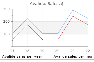
Avalide dosages: 162.5 mg
Avalide packs: 30 pills, 60 pills, 90 pills, 120 pills, 180 pills, 270 pills

Generic 162.5 mg avalide mastercardThe surgical method depends on the location of the lesion blood pressure 80 over 40 avalide 162.5 mg buy discount line, the necessity for mind rest blood pressure chart for child cheap 162.5 mg avalide amex, and whether or not exposure would require mind resection heart attack cafe menu avalide 162.5 mg buy with visa. Patient positioning usually is dependent upon the situation and surgical strategy to the tumor (Table 1 blood pressure erratic cheap avalide 162.5 mg without a prescription. Intraaxial (Within-the-Brain Parenchyma) vs Extraaxial (Outside-theBrain Parenchyma) Table 1. Common Patient Positions Based on Tumor Location Several kinds of incisions are used for these procedures. Linear incisions can be used to resect small tumors over the convexity or when using a midline method to the posterior fossa and often have the benefit of a more fast wound closure. After the skull is exposed, burr holes are made with a drill, and the bone flap is reduce with the craniotome. Some surgeons routinely use a free-bone flap, by which the bone is completely eliminated and stored for the duration of the case. Other surgeons flip an osteoplastic flap, where the bone is left attached to muscle and/or pericranium to keep it partially vascularized. After the bone is removed, a quantity of small holes are drilled close to the sting of the craniotomy. This helps prevent blood from accumulating in the epidural house throughout the rest of the case. The methodology of dural opening generally is based on the scale of the bone opening and its proximity to venous sinuses. The surgeon then proceeds with tumor removal if it is on the floor or with mind retraction/resection if the tumor is deep to the floor. The bone flap is replaced and fixated, the galea is closed with sutures, and the pores and skin incision is closed with staples. Such monitoring is often used for tumors of the brain stem and cranium base, or when resecting tumors in critical locations. Image-guided navigation entails using a computerized workstation that may track the position of particular instruments earlier than and through the operation. This permits the surgeon to: (a) plan acceptable skin and bone openings, (b) select an optimum trajectory to the tumor, and (c) achieve a volumetric resection of the tumor. Data from the operative microscope can be integrated into the navigation system to aid in tumor dissection. Mild hypothermia is often utilized when resecting tumors in eloquent areas of the brain because hypothermia has been shown to present neuroprotection. Lumbar drainage typically is used for tumors within the cerebellopontine angle, within the pineal region, or tumors associated with vital edema. Awake craniotomies usually are reserved for tumors adjoining to or within the speech areas of the dominant cortex. Patients are sedated throughout cranial opening and are awakened as soon as the dura is open. Direct cortical stimulation is carried out to determine the relationship between the tumor and the speech facilities. The surgeon then maps out eloquent and noneloquent areas of the brain prior to starting the tumor resection. Interstitial chemotherapy or brachytherapy may be carried out after the tumor resection by implanting chemotherapy impregnated wafers or radioactive seeds within the resection cavity. In addition, a balloon tip catheter can be placed in the resection bed and injected with radioactive material at a later date. Variant procedure or approaches: Patient place varies depending on tumor location and surgeon desire (Table 1. Tumors on the convexity, or surface, of the mind could require minimal mind relaxation or exposure. Deep tumors, or tumors across the mind stem or skull base, might require substantial mind relaxation and retraction for optimal publicity. For deep tumors or tumors within or adjacent to crucial structures, the operating microscope is commonly used. Large vascular tumors could bear preoperative embolization of the tumor blood supply to decrease intraoperative blood loss. Discuss surgical, approach, positioning, tumor type, and potential for blood loss with surgeon. Chang Description: Tumors of the cranium fall into the classification of different bony tumors. Examples of forms of cranium tumors often requiring surgery include eosinophilic granuloma, histiocytosis, hemangioma, osteoma, epidermoid, dermoid, metastases, osteosarcoma, fibrous dysplasia, chordoma, chondrosarcoma, and meningioma. These tumors could occur anywhere on the cranium, except chordoma and chondrosarcoma, which normally arise from the cranium base. For instance, the sitting or inclined position usually is used for tumors within the occipital or suboccipital areas, and the supine position is used for frontal, temporal, or parietal tumors. Some surgeons choose the lateral place for temporal or parietal bone lesions and the inclined position for occipital and some suboccipital bone lesions. Then the affected skull is removed by slicing a bone flap around the tumor with the craniotome. In a suboccipital or posterior fossa craniectomy, the bone often is eliminated piecemeal with both a drill or a series of rongeurs. The surgeon may elect to perform a cranioplasty to cover the defect, depending on the dimensions and site. The defect may be repaired utilizing quite so much of strategies, which embody using a polymer, such as methylmethacrylate, or a bone cement, corresponding to hydroxyapatite. In addition, autologous bone could also be harvested from another location on the skull or one other site (hip or rib) for reconstruction. After the tumor is eliminated and the reconstruction is accomplished, the skin incision is closed. Occasionally, the cranium tumor might be approached intracranially, if its location favors that strategy. Examples embody tumors of the petrous portion of the temporal bone, fibrous dysplasia involving the optic canal, or cranium base tumors, corresponding to chordomas and chondrosarcomas. Chang Description: Head injuries occasionally require emergent surgical procedures to evacuate mass lesions or debride contused or contaminated brain. Often the complete head is shaved and placed in a headrest (pins, suction cups, or horseshoe). If the C-spine has not been cleared, the patient could additionally be placed in a lateral position to reduce neck involvement. For a standard trauma craniotomy, the scalp incision starts anterior to the tragus and continues superiorly in a question-mark sort path, ending in the frontal area. Gentle warm irrigation may be used to assist in elimination of an acute subdural hematoma. For penetrating head accidents or depressed cranium fractures, the wound is debrided, international our bodies are eliminated, bleeding is managed, and the dura is repaired if lacerated. Depending on the injury and the presence of mind swelling, it might be essential to shut the dura with autologous pericranium, or fascia lata. Alternatively, the dura may be left open, or a number of relaxing incisions could also be made within the dura to permit for brain swelling. The surgeon can typically repair and reconstruct a depressed skull fracture after removing a bone flap that surrounds the fracture. In addition, the bone flap may be overlooked to compensate for mind swelling and replaced later. Typically the supraorbital, auriculotemporal, lesser occipital and higher occipital nerves are blocked close to their scalp origins. In an acute subdural hematoma: A: burr gap is made, followed by craniectomy, and the incision is extended upward to form a big question mark, the medial extent of which follows the midline. The anterior burr gap is placed above the frontal sinus (the measurement of which could be estimated from preop radiographs). C: the dura can be opened with a Y- or X-shaped incision, with a flap being primarily based on the superior sagittal sinus.
162.5 mg avalide discount fast deliveryObturator nerve block with native anesthetic can be used as a diagnostic device throughout differential neural blockade on an anatomic foundation within the evaluation of hip pain arrhythmia specialist discount avalide 162.5 mg fast delivery. If destruction of the obturator nerve is being thought-about heart attack jaw pain right side generic avalide 162.5 mg without prescription, this technique is useful as a prognostic indicator of the degree of motor and sensory impairment that the affected person may expertise pulse pressure decrease avalide 162.5 mg purchase. Obturator nerve block with native anesthetic may be used to palliate acute pain emergencies pulse pressure pediatrics buy avalide 162.5 mg, including postoperative ache reduction, while ready for pharmacologic methods to turn out to be efficient. Obturator nerve block with local anesthetic can also be useful in the administration of hip adductor spasm, which might make perineal care or urinary catheterization troublesome. Obturator nerve block with native anesthetic and steroid is also helpful within the remedy of persistent hip pain when the pain is thought to be secondary to irritation or entrapment of the obturator nerve. A point 1 inch lateral and 1 inch inferior to the pubic tubercle is then identified and prepared with antiseptic resolution. The needle is superior approximately 3/4�1 inch deeper to place the needle tip in the obturator canal. Infection within the space of the obturator nerve represents a contraindication to obturator nerve block. Neurolytic block with small portions of phenol in glycerin or by cryoneurolysis or radiofrequency lesioning has been proven to present long-term aid for patients affected by continual pain secondary to trauma or tumor involving the hip joint in whom extra conservative therapies have been ineffectual. Destruction of the obturator nerve can also be helpful within the palliation of hip adductor spasm after spinal wire harm or stroke that limits the flexibility to present perineal care or allow sexual activity or urinary catheterization. Electromyography and magnetic resonance imaging of the lumbar plexus are indicated on this patient inhabitants to help rule out different causes of hip ache, including malignancy invading the lumbar plexus or epidural or vertebral metastatic illness at L2-L3-L4. The femoral nerve is derived from the posterior branches of the L2, L3, and L4 nerve roots. The femoral nerve is simply lateral to the femoral artery because it passes beneath the inguinal ligament and is enclosed with the femoral artery and vein inside the femoral sheath. The nerve gives off motor fibers to the sartorius, quadriceps femoris, and pectineus muscular tissues. The proper femoral nerve was injured throughout cardiac catheterization difficult by a retroperitoneal hematoma. The method can also be useful to present surgical anesthesia for the decrease extremity when combined with lateral femoral cutaneous, sciatic, and obturator nerve block or lumbar plexus block. Femoral nerve block with local anesthetic can be used diagnostically during differential neural blockade on an anatomic basis within the evaluation of lower extremity pain. If destruction of the femoral nerve is being thought of, this system is helpful as a prognostic indicator of the degree of motor and sensory impairment that the patient might expertise. Femoral nerve block with local anesthetic may be used to palliate acute pain emergencies, together with femoral neck and shaft fractures, and for postoperative pain aid whereas ready for pharmacologic methods to become efficient. Femoral nerve block with local anesthetic and steroid can be indicated in the palliation of pain and motor dysfunction related to diabetic femoral neuropathy. Local an infection involving the realm of the femoral nerve block can be a contraindication to the efficiency of femoral nerve block. A point simply lateral to the pulsations of the femoral artery and just inferior to the inguinal ligament is then identified and ready with antiseptic solution. If no persistent paresthesia is present and after cautious aspiration, 15�18 ml of 1. Subsequent every day nerve blocks are performed in an identical method, substituting 40 mg of methylprednisolone for the preliminary 80-mg dose. Because paresthesia is elicited with this technique, needle-induced trauma to the femoral nerve remains a risk. By advancing the needle slowly after which withdrawing the needle slightly away from the nerve, needle-induced trauma to the femoral nerve can be avoided. This approach is very helpful within the emergency division to present rapid relief for those sufferers affected by fractures of the femoral neck and shaft. Careful preblock neurologic assessment is necessary to avoid later attribution of pre-existing neurologic deficits to the femoral nerve block. It should be remembered that the most common explanation for pain radiating into the lower extremity is a herniated lumbar disc or nerve impingement secondary to degenerative arthritis of the backbone, not issues involving the femoral nerve per se. Electromyography and magnetic resonance imaging of the lumbar backbone, combined with the medical historical past and bodily examination, assist type out the etiology of femoral ache. The saphenous nerve is derived primarily from the fibers of the L3 and L4 nerve roots. The saphenous nerve is subject to trauma or compression anyplace along its course. The nerve is frequently traumatized during vein harvest procedures for coronary artery bypass grafting procedures. The saphenous nerve is also subject to compression as it passes over the medial condyle of the femur. The approach can be useful to provide surgical anesthesia for the distal decrease extremity when combined with tibial and common peroneal nerve block or lumbar plexus block. Saphenous nerve block on the knee with native anesthetic can be used diagnostically throughout differential neural blockade on an anatomic foundation within the evaluation of decrease extremity ache. If destruction of the saphenous nerve is being thought-about, this system is helpful as a prognostic indicator of the diploma of motor and sensory impairment that the patient might experience. Saphenous nerve block on the knee with local anesthetic could also be used to palliate acute ache emergencies, together with distal decrease extremity fractures and postoperative ache reduction, when combined with the beforehand talked about blocks while Medial epicondyle Femur waiting for pharmacologic methods to turn into effective. Saphenous nerve block at the knee with local anesthetic and steroid can also be indicated in the palliation of pain and motor dysfunction associated with diabetic neuropathy. Local infection involving the area of the saphenous nerve block can be a contraindication to the efficiency of saphenous nerve block. A level simply in front of the posterior edge of the medial condyle is then recognized and ready with antiseptic solution. Somatic Blocks of the Lower Extremity 475 redirected slightly extra anteriorly till a paresthesia is obtained. Once paresthesia is elicited within the distribution of the saphenous nerve, the needle is withdrawn 1 mm and the affected person is noticed to rule out any persistent paresthesia. Water-soluble distinction medium may be added to the local anesthetic to verify applicable needle placement. Because this method elicits a paresthesia, needle-induced trauma to the saphenous nerve stays attainable. By advancing the needle slowly and withdrawing the needle barely away from the nerve prior to injection, one can avoid needle-induced trauma to the saphenous nerve. Careful preblock neurologic evaluation is important to avoid the later attribution of pre-existing neurologic deficits to the saphenous nerve block on the knee. Compressive neuropathy of the saphenous nerve at the knee typically happens in musicians who play the cello. It must be remembered that the most common cause of ache radiating into the decrease extremity is herniated lumbar disc or nerve impingement secondary to degenerative arthritis of the backbone, not disorders involving the saphenous nerve per se. Other ache syndromes that might be confused with saphenous nerve entrapment embody lesions either above the origin of the saphenous nerve, corresponding to lesions of the femoral nerve, or lesions of the saphenous nerve at the ankle. The posterior femoral cutaneous department (S1-S3) innervates the posterior aspect of the thigh. Blood vessels accompanying the sciatic nerve at this point are the sciatic artery, a branch of the inferior gluteal artery, and the inferior gluteal veins. Therefore, a sciatic nerve block is normally utilized in mixture with a femoral nerve block, which anesthetizes the complete lower limb. For surgery of the ankle and foot, a sciatic nerve block at the side of a saphenous nerve block is indicated. For surgery and analgesia of the knee, the sciatic nerve block must be utilized in combination with blocks of the femoral, obturator, and lateral femoral cutaneous nerves. There are, nonetheless, risks associated with the long-term placement of epidural catheters. Therefore, using a new strategy of catheter placement on the sciatic nerve can present an alternative approach for pain relief. It can remove the danger of epidural abscess, hematoma formation, and catheter erosion of the dura. The affected limb can be individually handled without numbing or weakening the contralateral limb.
Diseases - Paruresis
- Syphilis
- LyP (lymphomatoid papulosis)
- Acrocephaly pulmonary stenosis mental retardation
- 48, XXYY syndrome
- Seizures benign familial neonatal recessive form
- Laryngeal abductor paralysis mental retardation
- Polymorphic macular degeneration
- Carnitine-acylcarnitine translocase deficiency
Avalide 162.5 mg buy mastercardComplications Complications of retrogasserian glycerol injection are paresthesia blood pressure control chart avalide 162.5 mg purchase visa, dysesthesia blood pressure 50 order avalide 162.5 mg amex, anesthesia dolorosa heart attack young square generic avalide 162.5 mg without a prescription, corneal hypoesthesia or anesthesia hypertension treatment 162.5 mg avalide fast delivery, diminished corneal reflex, keratitis, and masticatory weak point. Depending on the lateral rotation of the C-arm, the foramen ovale visualization can move extra medially toward the maxilla. During this injection, severe headache or dysesthesia may happen, and the affected person must be warned about this end result prior to the injection. Some sufferers could get benefit instantly, whereas some sufferers may experience relief within the subsequent 2 weeks. Apart from different strategies like glycerol, neurolytic solution injection, or balloon compression, the lesion is more exact with radiofrequency lesioning and must be limited to the affected nerve. Thus, through the sensorial stimulation the patient should be awake sufficient to respond to the take a look at with sensorial stimulation for proper localization of the tip of the electrode. This is also a method of verifying that the needle is passed by way of the foramen ovale and is on the retrogasserian rootlets. If the first and second divisions are affected, there ought to be no motor response. If the needle is correctly situated, there might be a tingling-like sensation or electric-like paresthesias within the innervation of that department within the face. However, it must be kept in thoughts that there may be residual sensorial deficits from earlier lesioning. Lesioning Several kinds of electrodes could also be used for lesioning, such as cordotomy-type electrodes and trigeminal electrodes with the Tew needle and the Racz-Finch curvedblunt needle. Lesion Parameters and its retrogasserian rootlets lie on a airplane running from a superomedial to inferolateral direction. After stimulation is completed, the doctor ought to again rule out if the needle is in a vessel or not. If blood continues to be aspirated, the procedure must be terminated and a second attempt ought to be made one other day. When the lesion is induced, examine the bilateral corneal reflex and ache sensitivity in the neuralgic and contralateral zones. A fourth lesion could also be assessed at 75�C if ache involves two branches of the V cranial nerve. The affected person is instructed to look forward to swelling of the face and to put ice on the face to cut back any swelling that will happen. Note the draping of the affected person with the world of entry exposed and an O2 cannula in place for trigeminal ganglion radiofrequency. Some authors prefer to do the lesioning on an outpatient basis, and a few hospitalize the patient for a day. The patient ought to be monitored for an additional month to determine if unwanted facet effects appear. The process should be accomplished with important sign monitoring because bradycardia and hypertension could also be noticed. Initially, an angiocatheter is launched at the entry site towards the foramen ovale. If more than one department of the trigeminal nerve is affected, a number of lesions by repositioning of the needle should be performed. After every repositioning, the stimulation check should be repeated to search paresthesia at the desired website. Note the curved-blunt Racz-Finch radiofrequency needle getting into the foramen ovale in its lateral facet. Complications Significant masseter weak spot is a common complication, especially in the preliminary interval. Hypoesthesia, dysesthesia, anesthesia dolorosa, balloon failure, and hematoma on the cheek may be noticed (Table 6-2). In chosen collection, Taha and Tew12 compared the outcomes and issues of percutaneous strategies. Retrobulbar Hematoma and Hematoma in Cheek If the needle is superior to the retrobulbar space, retrobulbar hematoma could develop. It is lowest for balloon compres- 86 Head and Neck conservative strategies without any sequelae. Compression over the attention stops the bleeding, and the swelling subsides during the following days. Compression over the cheek by cold pack after the needle is withdrawn may be useful. Misplacement of needles into incorrect cranium base foramina can result in vascular injury and secondary hypertension that, in turn, can result in bleeding. In case of deep needle placement, one can enter the brain stem and cause hemorrhage. Inadvertent injection of therapeutic agents into this cul-de-sac can spread to different intracranial buildings, producing profound and speedy lack of consciousness and collapse. This is obviously an eminently reversible scenario when local anesthetic brokers are used, however in the event that such a catastrophe occurred with neurolytic brokers, inadvertent neurolysis of adjacent cranial nerves may happen. Irritation of the dura might trigger persistent headache, and in some sufferers, nausea and vomiting lasting for days can also be noticed. If blood is aspirated, the needle ought to be replaced, and if bleeding continues, the process should be stopped. The endpoint is reached when the specified division of the trigeminal nerve has turn into barely analgesic however not anesthetic. Usually at about 70�C, analgesia occurs and additional coagulations are made on the similar temperature till some analgesia is produced within the required division. At this stage, the time for each coagulation can be elevated or decreased; nevertheless, if the temperature is increased with out first trying further time, anesthesia will abruptly develop. Sequential throbbing of the cannula might sometimes be observed through the early seconds of the lesion. To prevent hematoma in the cheek, ice compression after the needle is withdrawn must be carried out in each occasion. Weakness of the homolateral masseter muscle might occur in the course of the postoperative interval. Because of the following analgesia of the conjunctiva, the eye have to be protected against persistent inflammatory processes that might go undetected because of the altered sensation. Protective spectacles with aspect shields can also help cut back the introduction of foreign bodies into the numb eye. Another problem with long-term hemifacial analgesia is saliva dribbling from the anesthetized half of the mouth; this can sometimes be alleviated by an antisialagogue similar to diphenhydramine, 25 mg tid. Shorter period of ache relief, higher recurrence charges, and growth of fibrosis on the foramen ovale are the primary disadvantages. Slight sensory deficit and Somatic Blocks 87 average price of recurrence could additionally be the benefits of gasserian ganglion compression. In one other research, technical failure for glycerol was reported to be as excessive as 15%. The highest price of recurrence is 54% for glycerol rhizotomy, with a imply follow-up of four years. Fibrosis could develop at the entrance of foramen ovale, enhancing further injections. All these techniques are much less morbid and more price effective than open surgical techniques. However, each technique should be applied in precise indications and in well-equipped facilities with experienced personnel. Comparison of Techniques In conclusion, the preliminary success fee with all three approaches is similar-91�93%. A variety of transient cranial nerve deficits had been also seen with a 2% probability of permanent ipsilateral hearing loss. However, far more regularly the indications are persistent, debilitating painful situations. Clearly, the use of fluoroscopy and extra training led to higher outcome and reduction of probably devastating problems.
Avalide 162.5 mg cheapCompletion of virus meeting leads to displacement of Hsc70 from virions (Florin et al heart attack would feel like a heart attack avalide 162.5 mg order amex. Virus like particles blood pressure jumps up cheap avalide 162.5 mg without prescription, however may be assembled by expression of L1 alone blood pressure chart printable generic 162.5 mg avalide with amex, the L2 protein is thought to enhance packaging and infectivity (Stauffer et al blood pressure medication for diabetics buy cheap avalide 162.5 mg on line. L2 interacts with L1 pentamers by way of the hydrophobic region in its C-terminus (Finnen et al. The interplay between capsomeres is mediated by the C-terminus of the L1 protein (Modis et al. Papillomavirus virions bear a really lengthy assembly course of inside tissue which is dependent on disulfide bond formation (Conway and Meyers 2009). Ultimately, virus launch requires environment friendly escape from the cornified envelope on the cell surface. Thus, it represents a by-product of viral an infection which will confer a selective benefit to the host cell without any obvious benefit to the virus (Pett and Coleman 2007; Jeon et al. Disruption of the viral genome also dissociates viral early (E) gene transcription from the viral early polyadenylation signal. Using host poly(A) signals, increases the stability of virus�host fusion transcripts with a longer half life (Jeon and Lambert 1995). In general, integration results in increased expression and stability of transcripts encoding the E6 and E7 proteins, along with the disruption of E2 protein. The overall differentiation-dependent papillomaviral gene expression noticed in vivo and in vitro involves promoter repression within the decrease strata and activation in the upper, differentiated strata. In contrast, malignant cervical most cancers cells retained the ability to express viral oncogenes following grafting (including basal layers), suggesting that mechanisms of viral transcriptional silencing were now not functional in these cells. These new insights point out earlier assumption that cervical neoplastic development occurs by way of integrant-only cells outgrowing episome-only cells is prone to be oversimplified. Progression of precursor lesions to cervical malignancy is characterised by a complex interaction between viral and host occasions. Progression of low-grade illness represents a breakdown of host controls that usually inhibit expression of episomal E6/E7 in the proliferating basal cells of the cervical squamous epithelium. Late events are host genomic modifications which would possibly be associated with acquisition of the invasive phenotype. Progression of high-grade illness is characterized by clonal enlargement of cells expressing E6 and E7 at an elevated degree, with consequent highlevel genomic instability and acquisition of mutations (Pett et al. Other factors, such as secondary infection with other pathogens, may trigger integrant selection by activating innate immune mechanisms that result in episome clearance (See Chap. Increased acetylation, particularly of the late promoter area means that altered chromatin construction might have been responsible, a minimum of in part, for the transcriptional deregulation in episomeassociated neoplastic progression. The L1 major capsid protein of human papillomavirus type 11 recombinant viruslike particles interacts with heparin and cell-surface glycosaminoglycans on human keratinocytes. Biology of the syndecans: a family of transmembrane heparan sulfate proteoglycans. Different heparan sulfate proteoglycans serve as mobile receptors for human papillomaviruses. Role of heparan sulfate in attachment to and infection of the murine feminine genital tract by human papillomavirus. Inhibition of switch to secondary receptors by heparan sulfate-binding drug or antibody induces noninfectious uptake of human papillomavirus. Mechanisms of human papillomavirus kind sixteen neutralization by l2 cross-neutralizing and l1 type-specific antibodies. Keratinocytesecreted laminin 5 can operate as a transient receptor for human papillomaviruses by binding virions and transferring them to adjacent cells. Human papillomaviruses bind a basal extracellular matrix component secreted by keratinocytes which is distinct from a membrane-associated receptor. Human papillomavirus type 31 makes use of a caveolin 1- and dynamin 2-mediated entry pathway for infection of human keratinocytes. Human papillomavirus kind sixteen infection of human keratinocytes requires clathrin and caveolin-1 and is brefeldin a sensitive. Analysis of the infectious entry pathway of human papillomavirus type 33 pseudovirions. Fusion of influenza virus in an intracellular acidic compartment measured by fluorescence dequenching. Caveolin-1-dependent infectious entry of human papillomavirus sort 31 in human keratinocytes proceeds to the endosomal pathway for pH-dependent uncoating. Interaction of human papillomavirus kind 16 L2 with mobile proteins: identification of novel nuclear body-associated proteins. Cleavage of the papillomavirus minor capsid protein, L2, at a furin consensus site is critical for infection. Identification of a dynein interacting area in the papillomavirus minor capsid protein l2. Identification of the dynein gentle chains required for human papillomavirus an infection. Amplification of human papillomavirus genomes in vitro is dependent on epithelial differentiation. Targeting the E1 replication protein to the papillomavirus origin of replication by advanced formation with the E2 transactivator. Segregation of viral plasmids is dependent upon tethering to chromosomes and is regulated by phosphorylation. Analysis of chromatin attachment and partitioning functions of bovine papillomavirus kind 1 E2 protein. The mitotic chromosome binding exercise of the papillomavirus E2 protein correlates with interplay with the mobile chromosomal protein, Brd4. ChlR1 is required for loading papillomavirus E2 onto mitotic chromosomes and viral genome upkeep. In vitro synthesis of oncogenic human papillomaviruses requires episomal genomes for differentiation-dependent late expression. The E8E2C protein, a adverse regulator of viral transcription and replication, is required for extrachromosomal maintenance of human papillomavirus kind 31 in keratinocytes. The E8�E2 gene product of human papillomavirus type 16 represses early transcription and replication however is dispensable for viral plasmid persistence in keratinocytes. Human papillomavirus sort 31 oncoproteins E6 and E7 are required for the maintenance of episomes in the course of the viral life cycle in normal human keratinocytes. Growth and differentiation of human papillomavirus kind 31b optimistic human cervical cell traces. Human papillomavirus type 31b E1 and E2 transcript expression correlates with vegetative viral genome amplification. The human papillomavirus sort sixteen E7 oncogene is required for the productive stage of the viral life cycle. Conditionally activated E7 proteins of high-risk and low-risk human papillomaviruses induce S phase in postmitotic, differentiated human keratinocytes. Life cycle heterogeneity in animal models of human papillomavirus-associated illness. Human papillomavirus oncoproteins alter differentiation-dependent cell cycle exit on suspension in semisolid medium. Temporal usage of multiple promoters through the life cycle of human papillomavirus sort 31b. Characterization of late gene transcripts expressed during vegetative replication of human papillomavirus kind 31b. Human papillomavirus kind 31 E5 protein supports cell cycle development and prompts late viral functions upon epithelial differentiation. The viral E4 protein is required for the completion of the cottontail rabbit papillomavirus productive cycle in vivo. Role of the E1�E4 protein in the differentiationdependent life cycle of human papillomavirus sort 31. A extremely environment friendly system to produce infectious human papillomavirus: Elucidation of pure virus-host interactions.
Avalide 162.5 mg buy otcThe wrist joint is innervated primarily by the deep branch of the ulnar nerve pulse pressure 29 discount avalide 162.5 mg otc, as properly as the anterior and posterior interosseous nerves blood pressure numbers low buy avalide 162.5 mg fast delivery. Anteriorly pre hypertension nursing diagnosis avalide 162.5 mg generic line, the wrist is bounded by the flexor tendons and the median and ulnar nerve arteria zigomatica avalide 162.5 mg mastercard. Medial to the joint runs the dorsal branch of the ulnar nerve, which is regularly broken when the distal ulna is fractured. Osteoarthritis of the joint is the commonest form of arthritis that leads to wrist joint ache. Less frequent causes of arthritis-induced wrist pain embrace the collagen vascular diseases, an infection, and Lyme disease. Acute infectious arthritis will normally be accompanied by important systemic symptoms including fever and malaise and should be simply acknowledged by the astute clinician and treated appropriately with tradition and antibiotics, somewhat than injection remedy. The collagen vascular illnesses will typically current as a polyarthropathy quite than a monoarthropathy restricted to the wrist joint, though wrist ache secondary to collagen vascular illness responds exceedingly well to the intraarticular injection technique described beneath. The majority of patients presenting with wrist pain secondary to osteoarthritis and post-traumatic arthritis pain will current with the complaint of pain, which is localized around the wrist and hand. In addition to the abovementioned pain, patients suffering from arthritis of the wrist joint will usually experience a gradual lower in functional capacity with lowering wrist vary of motion making simple everyday duties corresponding to utilizing a computer keyboard, holding a coffee cup, or turning a doorknob overhead fairly tough. Joint Blocks of the Upper Extremity 465 disuse, muscle wasting may happen and an adhesive capsulitis with subsequent ankylosis may develop. If resistance is encountered, the needle is probably in a ligament or tendon and should be advanced slightly into the joint space till the injection proceeds with out significant resistance. After sterile preparation of skin overlying the dorsal joint, the midcarpus proximal to the indentation of the capitate bone is recognized. Just proximal to the capitate bone is an indentation, which permits quick access to the wrist joint. If bone is encountered, the needle is withdrawn into the subcutaneous tissues and redirected superiorly. Slight irregularity of the distinction agent is seen in the radial aspect of the radiocarpal joint. Approximately 25% of sufferers will complain of a transient enhance in pain following intra-articular injection of the wrist joint and should be warned of such. Coexistent bursitis and tendonitis can also contribute to wrist ache and should require extra treatment with more localized injection of native anesthetic and depot steroid. This method is a protected process if cautious attention is paid to the clinically related anatomy within the areas to be injected. Care have to be taken to use sterile approach to keep away from an infection, as well as the usage of common precautions to keep away from threat to the operator. The incidence of ecchymosis and hematoma formation can be decreased if strain is placed on the injection web site instantly following injection. The use of bodily modalities including local warmth, in addition to gentle range of motion exercises, ought to be introduced sev- eral days after the patient undergoes this injection method for wrist ache. Hydrocortisone and cortisone injected into arthritic joints: comparative effects of and use of hydrocortisone as a local antiarthritic agent. In fact, shortly after Halsted and Hall launched the concept of the nerve block in December of 1884, Stillman reported the restore of a congenital foot abnormality beneath tibial nerve block carried out by Halstead. This landmark surgical procedure was performed on April 30, 1885, and it endlessly changed the widespread perception that "etherization" with all of its attendant dangers was the one option to keep away from the ache of surgical procedure. The ensuing years witnessed an explosion in analysis on the utilization of nerve block to provide anesthesia for lower extremity surgery. One must surmise that this search for higher ways to render the decrease extremity insensate would have continued unabated however for the speedy acceptance of spinal anesthesia by Corning in the years following its introduction in 1885. By 1900, spinal anesthesia had gained extensive acceptance and had supplanted common anesthesia and regional blocks as the preferred form of anesthesia for the decrease extremity due to its ease of efficiency and reliability. Many surgical and later anesthesia coaching programs dropped regional nerve blocks of the decrease extremity from their curricula in favor of spinal anesthesia. Mirroring the waning interest in regional anesthesia generally in each the operating room and obstetrical arenas, it was the introduction of the safer amide native anesthetics and specifically the introduction of the longer-acting amide-type medication similar to bupivacaine that led to a renewed interest in regional blockade of the somatic nerves of the lower extremity, not for surgical anesthesia, however for use in postoperative ache aid. Today, the blocks described beneath represent the usual of care for postoperative ache aid in most busy surgical facilities and hospitals. The nerves lie in entrance of the transverse processes of their respective vertebrae; as they course inferolaterally, they divide into a number of peripheral nerves. The obturator nerve receives fibers from L2-L4, and the femoral nerve is made up of fibers from L2-L4. This variability signifies that differential neural blockade on an anatomic basis have to be interpreted with warning. The rationale behind lumbar plexus block using the Winnie 3-in-1 technique is to block the three principle nerves that compose the lumbar plexus as they lie enclosed 467 468 Extremities Femoral nerve Lumbosacral trunk Inguinal ligament Obdurator nerve Lumbar plexus nerve block via the Winnie 3-in-1 method with native anesthetic could also be used to palliate acute ache emergencies, including groin and decrease extremity trauma or fracture, acute herpes zoster, and cancer pain, whereas ready for pharmacologic, surgical, and antiblastic therapies to turn out to be efficient. Lumbar plexus nerve block via the Winnie 3-in-1 approach with local anesthetic and steroid is also useful within the treatment of lumbar plexitis secondary to virus or diabetes. For most surgical and pain management applications, epidural or subarachnoid block is a greater different, though one ought to anticipate fewer cardiovascular changes with lumbar plexus block in contrast with epidural or subarachnoid methods. More selective methods corresponding to radiofrequency lesioning of specific lumbar paravertebral nerve roots may trigger less morbidity than lumbar plexus neurolysis. Local an infection involving the area of the lumbar plexus can additionally be a contraindication to the efficiency of lumbar plexus block. Note the course of the femoral nerve and the obturator nerve as it exits from the pelvis to the groin. Lumbar plexus nerve block through the Winnie 3-in-1 method is used primarily for surgical anesthesia of the decrease extremity. It is often used within the space of pain administration when treating pain secondary to inflammatory circumstances of the lumbar plexus or when tumor has invaded the tissues subserved by the lumbar plexus or the plexus itself. Lumbar plexus nerve block by way of the Winnie 3-in-1 method with native anesthetic is often used diagnostically throughout differential neural blockade on an anatomic foundation within the analysis of lower extremity and groin ache. If destruction of the lumbar plexus is being thought-about, this method is helpful as a prognostic indicator of the diploma of motor and sensory impairment that the affected person may experience. The inguinal ligament and the femoral artery on the facet to be blocked are recognized. At a point simply lateral to the femoral artery and slightly below the inguinal ligament, the pores and skin is ready with antiseptic answer. Somatic Blocks of the Lower Extremity Femoral nerve Inguinal ligament Femoral artery Femoral vein Iliopsoas muscle Pectineus muscle 469 sequelae. Post-block groin and again pain, in addition to ecchymosis and hematoma of the groin, happen usually enough that the affected person must be warned of such previous to beginning lumbar plexus block utilizing the Winnie 3-in-1 technique. It has the advantage over the psoas compartment approach in that it allows straightforward catheter placement for continuous infusions of native anesthetic. Unfortunately, many of the issues that can be carried out with lumbar plexus block may be accomplished more easily with epidural or spinal techniques, which may be more acceptable to the surgeon and pain specialist alike. Neurolytic block with small quantities of phenol in glycerin or with absolute alcohol has been proven to provide long-term relief for patients suffering from cancer-related ache in whom more conservative remedies have been ineffectual. As mentioned earlier, the proximity of the femoral artery and vein makes cautious attention to approach obligatory. The pain specialist should rigorously study the patient prior to performing lumbar plexus block using the Winnie 3-in-1 technique to determine pre-existing neural compromise which may subsequently be erroneously attributed to the block. Pressure must be applied beneath the needle to force the answer to flow cranially alongside the fascial airplane rather than distally into the leg. Subsequent day by day nerve blocks are carried out in an analogous method, substituting 40 mg of methylprednisolone for the preliminary 80-mg dose. As mentioned earlier, an intravenous catheter can be positioned into the fascial sheath to allow steady infusion of local anesthetic. Persistent paresthesia secondary to trauma to the femoral nerve has rarely been reported after this method. Although unusual, infection remains an ever-present risk, particularly within the immunocompromised most cancers affected person. The posterior department provides motor innervation to the deep hip adductors and an articular department to the posterior knee joint. The technique can be useful to provide surgical anesthesia for the decrease extremity when combined with lateral femoral 470 Extremities Lat.

162.5 mg avalide order overnight deliveryWith a suggestive Hx pulse pressure emt avalide 162.5 mg, a brand new murmur blood pressure 8060 order 162.5 mg avalide free shipping, or a pulse deficit blood pressure 5080 purchase avalide 162.5 mg amex, an enlarged mediastinal shadow on chest radiography should immediate additional diagnostic efforts arteria vesicalis generic avalide 162.5 mg overnight delivery. Through a median sternotomy, venous access is gained by way of the right atrium, and arterial inflow is equipped through a femoral artery or axillary artery. During a interval of circulatory arrest, antegrade or retrograde cerebral perfusion, the ascending aorta is opened and the tear localized. The restore is carried distally into the arch, if the complete dissection could be resected, and the distal aortic layers are reapproximated with a Teflon felt strip supporting the medial and adventitial layers. The distal graft anastomosis is then accomplished, the graft clamped, the bypass pump restarted, and systemic warming commenced. Proximally, the aortic root is reconstructed, again using Teflon felt to help the medial and adventitial layers and to resuspend the aortic valve, which could be salvaged in roughly 85% of circumstances. The heart is then cleared of air and the cross-clamp eliminated to permit reperfusion of the coronary circulation. After oversewing patent intercostal arteries, the medial and adventitial layers are buttressed with Teflon felt, and an interposition Dacron graft is sewn into place. Heparin reversal, decannulation, and closure are achieved within the usual manner. The three classification techniques of aortic dissection and the distribution of the intimal tear. Dorotta I, Kimball-Jones P, Applegate R: Deep hypothermia and circulatory arrest in adults. Fattori R, Mineo G, Di Eusano M: Acute sort B aortic dissection: present administration methods. Lips J, de Haan P, de Jager S, et al: the role of transcranial motor evoked potentials in predicting neurologic and histopathologic outcome after experimental spinal twine ischemia. Lu S, Sun X, Hong T, et al: Bilateral versus unilateral antegrade cerebral perfusion in arch reconstruction for aortic dissection. Olsson C, Thelin S, Stahle E, et al: Thoracic aortic aneurysm and dissection: rising prevalence and improved outcomes reported in a nationwide population-based research of greater than 14,000 cases from 1987 to 2002. Penco M, Paparoni S, Dagianti A: Usefulness of transesophageal echocardiography within the evaluation of aortic dissection. In addition, the blood provide to the spinal twine might arise from lumbar and/or intercostal vessels within the affected aortic section, producing critical cord ischemia throughout cross-clamping and postop paraplegia. Almost all thoracoabdominal aneurysm repairs are carried out via a thoracoabdominal incision. After opening the chest, the incision is prolonged across the costal cartilage onto the stomach. The diaphragm is radially incised to the aortic hiatus, and the retroperitoneal dissection aircraft established anterior to the psoas musculature. All intraabdominal contents, in addition to the left kidney, are mirrored anteriorly. Back-bleeding from patent intercostal, mesenteric, and renal vessels could be controlled by balloon catheters, and aggressive blood salvage with autotransfusion gadgets is necessary. The restore entails suturing a tube graft proximally to the divided aorta and then sewing islands of aortic tissue containing intercostal visceral vessels onto appropriate-sized holes in the side of the tube graft. This allows reperfusion of important intercostal, celiac axis, superior mesenteric, renal arteries, and finally, the distal aorta or iliac arteries. During the inclusion technique, the anterior renal fascia is opened and the kidney is mobilized, together with the upper stomach organs (on the left). After aortic cross-clamping, the aneurysm is opened and the repair performed from within the aneurysm, stitching on-lay patches of the intercostal, mesenteric, and renal vessels to openings created within the tube graft. This no-clamp approach allows cheap management of those very extensive aneurysms, but leads to an obligatory and ongoing blood loss by way of back-bleeding of visceral vessels till the anastomoses are full. Patients presenting electively for thoracotomy (descending aorta) may have a thoracic epidural catheter placed the evening before surgery. Kazama S, Masaki Y, Maruyama S, et al: Effect of altering cerebrospinal fluid strain on spinal twine blood flow. Ling E, Arrellano R: Systematic overview of the evidence supporting the utilization of cerebrospinal fluid drainage in thoracoabdominal aneurysm surgery for prevention of paraplegia. Although aortic aneurysms might involve the suprarenal aorta, the majority are infrarenal in origin and may lengthen into the iliac arteries. Most (> 95%) are asymptomatic and are found by the way throughout investigation of one other medical problem. Because of the related elevated danger for rupture as the aneurysm increases in measurement, most vascular surgeons advocate prophylactic restore for aneurysms > 5 cm in crosssection dimension. Endovascular stent-grafting of belly aortic aneurysm has emerged instead therapeutic approach. After publicity of the stomach aorta from the extent of the renal vein distally to the iliac arteries, the aorta is crossclamped, distally at first to stop atheroembolism, and then proximally. Graft origin is often from the infrarenal aorta, however may arise from the intramesenteric aorta or even the supraceliac aorta. Graft termination may be to the distal aorta above the bifurcation (tube graft), to the common or exterior iliac arteries (Y graft), or the femoral arteries. Immediately prior to cross-clamping, vasodilators are elevated to scale back afterload, which is considerably elevated with software of the aortic cross-clamp. The aorta is then incised, lumbar vessels oversewn, and the aorta transected to allow an interposition graft to be sewn into place. The retroperitoneal method has many advocates, as it could require less quantity intraop, may be related to much less temperature loss, and will result in a shorter interval of postop adynamic ileus. In a randomized, prospective research, nonetheless, no vital differences could probably be detected between these two approaches for blood loss or postop recovery time. Retroperitoneal strategy to the aorta: the incision is prolonged from the tip of the 11th rib or tenth intercostal house laterally toward the midhypogastrium. Nevertheless, hemodynamic monitoring is necessary to enable for rapid quantity shifts and to ensure sufficient preload and adequate afterload discount, particularly through the interval of aortic cross-clamping. Boccara G, Jaber S, Eliet J, et al: Monitoring of end-tidal carbon dioxide partial strain adjustments during infrarenal aortic cross-clamping: a non-invasive method to predict unclamping hypotension. Filinger M: Who should we operate on and how will we decide: predicting rupture and survival in patients with aortic aneurysm. Panaretou V Toufekzian L, Siafaka I, et al: Postoperative pulmonary perform, after open stomach aortic aneurysm repair in patients with persistent obstructive pulmonary disease: epidural versus intravenous analgesia. Patra P, Chaillou P, Bizouarn P: Intraoperative autotransfusion for restore of unruptured aneurysms of the infrarenal abdominal aorta. Sprung J, Abdelmalak B, Gottlieb A, et al: Analysis of risk components for myocardial infarction and cardiac mortality after main vascular surgery. Its use is predicated on the existence of enough influx to the level of the groin or femoral artery. Other needed parts embody an enough goal vessel, preferably in continuity with runoff into the plantar arch of the foot, and an adequate conduit, preferably autologous saphenous vein. Long-term patency rates of nonautologous conduits to the below-knee arteries are distinctly inferior to that of saphenous vein and ought to be avoided whenever possible. The operative repair usually entails incisions on the groin and distal bypass websites to expose the donor and recipient arteries and to harvest leg or arm venous conduit. An unobstructed influx source- usually the common femoral, superficial femoral, or deep femoral artery-is exposed in the groin. The goal distal artery, often on the level of the knee or under, can be approached by way of a medial incision. More distally, the peroneal and anterior tibial arteries can be approached laterally on the midtibial level. At the extent of the malleolus, each the dorsalis pedis and posterior tibial arteries can be revascularized. After management of donor and recipient vessels, an anatomic tunnel is created, and the bypass conduit (saphenous vein, prosthetic graft) is passed via its length. After administration of 10,000 U of heparin, the distal anastomosis is constructed first, followed by the proximal anastomosis. Heparin is then partially reversed and meticulous hemostasis obtained, and the injuries are closed.
Manggistan (Mangosteen). Avalide. - Dysentery, diarrhea, urinary tract infections (UTI), gonorrhea, thrush, tuberculosis, eczema, menstrual disorders, and other conditions.
- Are there safety concerns?
- How does Mangosteen work?
- Dosing considerations for Mangosteen.
- What is Mangosteen?
Source: http://www.rxlist.com/script/main/art.asp?articlekey=97027
Avalide 162.5 mg buy cheap on-lineSilastic tubing from the band is then connected to a port placed in the subcutaneous tissue blood pressure medication morning or evening generic avalide 162.5 mg on-line. An alternative open process is the biliopancreatic diversion with duodenal switch blood pressure and exercise 162.5 mg avalide cheap otc, which involves a longitudinal gastric resection with a proximal small bowel bypass hypertension 16070 order avalide 162.5 mg line. Another different open procedure is a sleeve gastrectomy heart attack 1d lyrics avalide 162.5 mg buy low price, the place a gastric tube is created from the gastroesophageal junction to the pylorus over a 36�F dilator. Both of these operations are gaining recognition within the bariatric surgery group. This procedure has been largely deserted due to the many associated complications, including cirrhosis, osteoporosis, kidney stones, and intractable diarrhea. Another alternative open procedure is a sleeve gastrectomy the place a gastric tube is created from the gastroesophageal junction to the pylorus over a 36�F dilator. A: In commonplace supine position, the atlantooccipital gap of morbidly overweight affected person is obliterated by fats, and access for laryngoscope is hindered by massive breasts. B: By elevating the shoulders and occiput in order that head is within the "sniffing" position, airway entry is greatly facilitated. Obesity, and size of publicity to weight problems, enhance the chance of hospital admission and lengthen hospital keep. Review a listing of all current drugs the patient is taking, including nonprescription urge for food suppressors and food plan drugs. A reduction in vitamin K ranges can increase the anticoagulation results of Coumadin (also see Anesthetic Considerations for Laparoscopic Bariatric Surgery, p. If using a combined anesthetic approach, placement of an epidural catheter must be achieved earlier than induction, with the affected person within the sitting place. A bilateral sensory block (using 5�7 mL 2% lidocaine) will assist affirm correct placement of the catheter within the epidural house. Verification of placement is particularly essential in this population as a outcome of regional anesthesia within the obese affected person could additionally be technically tougher. Perilla V, Sollazzi L, Bozza P, et al: the results of the reverse Trendelenburg place on respiratory mechanics and blood gases in morbidly obese sufferers throughout bariatric surgical procedure. Such tubes can be utilized for gastric decompression or for feeding, and so they could also be everlasting or temporary. Patients undergoing gastrostomy placement often have neurologic impairment that compromises their ability to handle oral secretions and will increase their threat of aspiration. The anterior wall of the abdomen is recognized, and two purse-string sutures are placed within the stomach around the web site where the tube will enter. The gastrostomy tube is launched through the abdominal wall directly over the meant site of entry into the stomach. A small hole is made in the abdomen within the heart of the purse-string sutures, the tube is introduced into the stomach, and the purse-strings are tied securely around the tube. The greater curvature of the stomach is recognized and a stapler positioned throughout a portion of this, creating a tube that arises from the principle physique of the stomach. The staple line may be oversewn, after which the end of the tube is brought by way of the belly wall and matured to the pores and skin as a small stoma. This allows for everlasting entry to the stomach with elimination of the tube between feedings and is beneficial in sufferers with long-term dependence on gastrostomy entry. The gastrostomy tube is introduced via the mouth and handed via the abdomen and abdominal wall from inside out. Previous gastric operations may make endoscopic placement tough or harmful, as could some obstructing lesions of the esophagus or pharynx. To avoid injury to the back wall of the abdomen, the anesthesiologist could additionally be requested to inject air forcefully into the stomach. Percutaneous gastrostomies often are placed in sufferers with superior malignancy and intestinal obstruction or inadequate oral intake, and in sufferers with neurologic impairment and issue eating. It is essential, therefore, to be familiar with the anatomy of the proximal duodenum in relation to the major and minor pancreatic duct orifices. A transverse opening permits one to shut the duodenotomy without tension; nonetheless, it must be made very precisely for the purpose of publicity. Care should be taken to avoid perforating the duodenum when performing a sphincterotomy. The appendix is then delivered via the wound; and the mesoappendix is clamped, cut, and ligated. The appendiceal stump may be invaginated into the wall of the cecum or left alone. In some instances it may be simpler to divide the bottom of the appendix before delivering the appendix into the wound. The wound ought to be left open and delicate drains used in circumstances of perforated appendix. In laparoscopic procedures, the appendix is usually transected with an endoscopic stapling system. Ectopic mucosa is current in roughly 50% of symptomatic patients, with gastric mucosa the most frequent. After entering the peritoneal cavity, the distal ileum, together with the diverticulum, is delivered into the wound. Following excision of the diverticulum, care should be taken to not narrow the bowel lumen during closure. If a prognosis can be made preop, a laparoscopic strategy may be used (see Laparoscopic Bowel Resection p. These sufferers are in danger for aspiration pneumonitis because of delayed gastric emptying, ileus, or frank bowel obstruction full stomach precautions are appropriate. Surgery for appendicitis is among the most typical nonobstetric procedures performed on the pregnant affected person (~1/1500 pregnancies). These patients often are extra sick on the time of diagnosis because early signs could additionally be attributed to being pregnant, and the gravid uterus could hinder an correct belly exam. Anesthesia management for the gravid appendicitis affected person mirrors that of the nongravid patient (full-stomach precautions) with consideration of the maternal physiologic modifications of being pregnant and the consequences of anesthesia on the fetus and uteroplacental perfusion (See Anesthetic Considerations for Cervical Cerclage, Obstetric Surgery, p. If systemic sepsis is absent, hydration is adequate, the patient is cooperative, and high abdominal exploration is unlikely, then regional anesthesia could additionally be considered for open procedures. An intestinal tube is either purse-stringed into the small bowel and introduced by way of the belly wall or the intestine itself is introduced to the exterior and customary into a stoma. After purse-stringing the tube in the bowel, the seromuscular layer of the jejunum is sutured over the tube for a distance of 3�4 cm before exiting through the stomach wall. The Brooke ileostomy is created by bringing a 2-inch section of ileum through an stomach wall stab wound. For instance, sure tubes are used for feeding, whereas others may be used for drainage or decompression. Many of those sufferers will have irregular protecting airway reflexes and are vulnerable to aspiration of gastric contents. Cingi A, Solmaz A, Attaqllah W, et al: Enterostomy closure site hernias: a scientific and ultrasonographic evaluation. Approximately 45 cm of small bowel are required for building of the pouch and valve. After suturing two limbs of the ileum collectively over a distance of 15 cm, the distal section is intussuscepted over itself to type the nipple valve. The pouch is then sutured closed and mounted beneath the abdominal wall stoma web site. The stoma is made flush with the pores and skin for cosmetic causes and left intubated for 1 month with a particular plastic catheter. The pouch remains decompressed for 1 month before intermittent catheterization is initiated. The continent ileostomy reservoir has been modified by Barnett to include the construction of an isoperistaltic valve with an intestinal collar around its base to stop deintussusception and valve prolapse. These procedures are typically carried out following a complete proctocolectomy or to replace typical ileostomies. After coming into the peritoneal cavity, the involved small bowel is delivered into the wound and the lesion resected between bowel clamps.
Generic 162.5 mg avalide fast deliveryA triple-antibiotic ointment is placed on the wound and is roofed by a bandage or different applicable dressing blood pressure medication causes cough generic avalide 162.5 mg overnight delivery. We have demonstrated in our laboratory that when methylprednisolone (Depo-Medrol) plus local anesthetic or triamcinolone (Aristocort) and native anesthetic are injected through a bacteriostatic filter blood pressure medication list a-z order 162.5 mg avalide amex, the filter screens out just about all of the steroid blood pressure medication hydralazine avalide 162.5 mg generic online. For the infusion pulse pressure chart purchase avalide 162.5 mg fast delivery, the affected person is placed within the lateral place with the painful aspect down and stays on this place for half-hour following the com- 416 Pelvis that the patient hold the world dry for forty eight hours after removing to decrease the possibility of an infection. Showering is permitted after this era, but immersion of the wound similar to in a bath or pool remedy must be avoided for a minimal of 7 to 10 days. This procedure is usually followed by important improvement in ache and motor perform. With improvement of ache, it may be very important provoke aggressive bodily remedy to enhance muscle energy and tone, which is usually decreased from lack of use secondary to pain. Because of the steroids used, a 3-month delay between procedures is critical, during which time the patient ought to be inspired to proceed intense bodily remedy. Initiation of neural flossing techniques, especially while the native anesthetic remains to be lively, offers a main opportunity to maximize the adhesiolysis process with the least discomfort to the affected person. One month of aquatic remedy adopted by aggressive, graded physical therapy and work hardening can additionally be really helpful. This is adopted by the injection of 10 ml of preservativefree saline, 1500 U of hyaluronidase spreading to L4 and L5, and at last the ten ml of 0. The contrast is spreading as a lot as L4, L5, and then to S1, evenly, almost like a Christmas tree appearance. The foot drop dramatically improved the following day on account of the decompression of the L4-L5, S1 nerve roots by dissection of the perineural area with the injected materials. Immediate monitoring is for acute effects of spinal local anesthetic and mechanical issues. The initial 4 hours should embody mattress rest or supervised strolling, as partial motor block may be current. Bladder perform and motor recovery ought to be current before the affected person is allowed to ambulate independently. Patients ought to be warned that perianal numbness may be transiently present, especially in patients with spinal stenosis as a outcome of a transient neuropraxia. A subdural block is commonly typified by a segmental motor block with a diffuse sensory block to the extent expected from a subarachnoid injection of local anesthetic. If blood is aspirated, the needle is first retracted caudad within the sacral canal till no blood can be aspirated. If that is unsuccessful, an attempt can be made to proceed with catheter placement into the correct website. Aspiration through this catheter should be unfavorable for blood, and lack of venous runoff ought to be confirmed with injection of contrast medium. The opposed results include bruising, transient hypotension, transient breathing problem, numbness of the extremities, bowel or bladder dysfunction, paralysis, infection, sexual dysfunction, and the likelihood that the catheter might shear. The most common idiosyncratic response occurring after intrathecal iohexol is headache, which occurs in approximately 18% of sufferers. Aseptic meningitis and neurologic disturbances have been reported as rare issues. In addition, allergic or anaphylactoid reactions could occur not often with nonionic brokers, however far less regularly than with their ionic predecessors. Ndosi and colleagues69 determined in a double-blinded, placebocontrolled trial that myelography with applicable concentrations of iohexol (240 mgI/ml) carried no more risk of significant reaction than a diagnostic lumbar puncture. The same examine demonstrated that iohexol concentrations of a hundred and eighty and 300 mgI/ml had been extra more probably to result in headache, dizziness, nausea, vomiting, and seizures. Insect hyaluronidase is an allergen in stinging-insect venoms and has a recognized homology to mammalian hyaluronidase. This usually outcomes from unintended intravascular administration of large doses of bupivacaine. Bupivacaine disassociates from sodium channels extra slowly than lidocaine throughout cardiac diastole; due to this fact, its effect is more Spinal Neuroaxial Procedures 417 pronounced and cumulative. Cases of aseptic and bacterial meningitis, as well as epidural abscess, have been reported rarely. They include cardiac, respiratory, or neurologic sequelae in approximately 10% of patients. Arthur and colleagues103 described a study at the seventh World Congress on Pain in which the lysis of adhesions approach was equivalent to the present technique in a randomly selected group of patients for a retrospective evaluate. A study by Devulder and coworkers104 in 1995 was based mostly on 34 sufferers in whom epidural adhesions have been suspected based on both magnetic resonance imaging or their history of back surgical procedure. In their protocol, during which hyaluronidase was not employed, an epidural catheter was placed through the sacral hiatus underneath fluoroscopy but without path towards the affected web site. The catheter was simply superior 10 cm into the epidural area, and 10 ml of contrast agent (10 hexol 240 mgI/ml) was injected. Injection of 20 ml of 2% lidocaine with 80 mg of methylprednisolone added was adopted by 10 ml of 10% hypertonic saline. The researchers noted a regression of adhesions in 14 of the 30 patients who had had defects. Seven of those sufferers reported marked improvement of their pain, defined as a visible analog scale rating of lower than 4 at 1 month. Only two of those patients reported this degree of enchancment at 3 months, and at 1 year this whole group of sufferers had undergone a unique remedy as a result of their ache returned. Only four of the patients with none enchancment of contrast spread reported marked pain aid at 1 month, two at 3 months; and one remained pain free at 1 12 months. Chi-square analysis of those information confirmed no statistically vital correlation between enhanced contrast unfold after the injections and a greater consequence. This process has been criticized for the dearth of guidance of the catheter tip into the lesion and demonstrates the importance of directing the catheter tip into the lesion. The idea of transforaminal needle and catheter technique was first presented by Michael Hammer. The borders of the lumbar foramen consist of the vertebral physique and disc anteriorly, the pedicles superiorly and inferiorly, and the side explicit processes posteriorly. Anterior to the nerve root, radicular vessels can be discovered to observe the nerve root into the epidural space. Using sterile preparation and method, the back is cleansed with a sterilizing resolution from just below the scapula to the decrease margin of the buttocks. Preparation of the lumbar area is suitable only if the higher lumbar area is the supply of the problem, without sciatic involvement. The fluoroscope is then oblique to the ipsilateral facet of the desired foramina to 15�20 levels. A caudal-cephalad rotation elongates the superior articular course of ("ear of the Scottie dog"). The tip of the ear or superior articular course of in a "gunbarrel" approach is marked on the skin. This spot is the pores and skin entry website, and local anesthetic is injected for pores and skin infiltration. Next, a lateral fluoroscopic view is obtained prior to additional introduction of the needle. To facilitate passage of the needle previous the articular process, the epidural needle is turned laterally to slide previous the bone and turned medially and slowly advanced until a "pop" is felt. The needle tip on a lateral view should be within the posterior side of the foramen. Occasionally, the epidural needle should be tilted on the hub laterally to help entry of the epidural catheter into the anterior epidural space. Aspiration should be negative earlier than 3 ml of iohexol (Omnipaque 240) radiographic contrast is injected. The contrast injection should present opening of the entered neuroforamen with contrast agent exiting along the path of the nerve root. When satisfactory contrast spread is seen, 6 ml of 1500 U of hyaluronidase in 10 ml of preservative-free regular saline are injected to facilitate the opening of different adhesions. During the injection, the contrast have to be seen spreading out via the neural foramen, and hyaluronidase has a big function in opening up the scarreddown epidural house. If all of the injection volume loculates throughout the epidural house with out spreading outdoors, it could characterize a big loculation hazard.
Generic avalide 162.5 mg visaIn a microscope we observe a shift in epithelial keratinization sample with hyperkeratinization and hyperplasia; epithelial atrophy from the dorsum of the tongue specimen; epithelial ulceration with lack of mucosa covering with exposure of underlying connective tissue hypertension benign 162.5 mg avalide discount mastercard, and medical leukoplakia with microscopic epithelial hyperplasia blood pressure medication ratings avalide 162.5 mg cheap without prescription. Other scientific modifications are odontogenic cervical cuffing attributable to Streptococci sp arrhythmia bradycardia discount 162.5 mg avalide with mastercard. In the oral pharynx Firmicutes is in an inverse correlation with Proteobacteria arteria femoralis generic avalide 162.5 mg with mastercard, which occurs extra often within the distal esophagus or mouth than within the saliva (Lemon et al. Moreover, presence of selective microbial populations within the pharynx and oral cavity are a product of a clearing mechanism from the digestive and respiratory tract. Part of this process involves direct inhalation of microbes from the surroundings to introduce new opportunistic organisms. These microbes can compete for specific niches and change microbe survival (Hooper et al. This is obvious in distinct phylum-level distribution patterns for ecologic niches (Mager et al. Therefore a pure dynamic in microbial populations can affect threat for varied cancers. It can also be the positioning for many necessary human pathogens, together with Streptococcus pneumoniae, Streptococcus pyogenes, Haemophilus influenzae, Neisseria meningitidis, Moraxella catarrhalis, and Staphylococcus aureus (Meurman and Uittamo 2008; Hooper et al. These bacterial households are capable of producing a posh microbial setting that manifest as abscesses/fistulous tract, granuloma, lympho-granuloma, or fibrosis/scarring in the mucosa. In the oral cavity microbial populated plaques or calculus formations reside in gingival cervical sulci or connect to odontogenic Poly-Microbial Interaction with Human Papilloma Virus Leading to Increased. In these anatomic niches bacterial populations proliferate to trigger clinical damage to epithelial mucosa, odontogenic buildings, and gingival surfaces (Robertson and Smith 2009; Gill and Scully 1990; Brook 2006; Meyer et al. A dependent phase related to a host inflammation is a consequence as this interplay continues to elaborate. Clinically we observe losses of normal mucosa operate, mucosal overlaying and a depressed host safety from microbes as abscesses and granulomas appear. Manifestations of this process within the epithelial mucosa would include modifications in mucosal histopathology similar to: elevated florid keratinization, presence of isolated dyskeratotic cells, elevated epithelial hyperplasia, mitotic activity in supra basal and basal areas, growth of rete pegs to type pseudo-epithelial hyperplasia with extension of rete pegs, and presence of acanthous in suprabasal and basal areas of the mucosa. Numbers of inflammatory effectors: Langerhan cells (tissue specific histiocytes), granulocytes, lymphocytes are also noticed to infiltrate into the epithelium. On occasion microabscesses that would indicate an acute inflammatory response to foreign-microbial protein are discerned in suprabasal regions. Keratinocyte change can also be recognized to embody hyperchromatism, nuclear-cytoplasmic ratio reversal, anaplasia, pleomorphism, dyskeratosis, and mitotic figures. Increasing severity for premalignant changes embody the fore-mentioned features with presence of weird mitoses, and micronuclei with heterochromatin debris in basal and supra basal nuclei. Identification of those options helps to affirm a lack of tight intracellular bridges, and a weakening of gap junction-adherens-desmosomal tight junctions. These epithelial intracellular attachment websites are important for permeability in the oral mucosa. Tight Junctions arrange epithelial polarization and establish an apico-lateral barrier for management of solute diffusion via the intracellular area (gate function). These tight junction regions also prohibit movement of lipids and membrane proteins between the apical keratinocyte area and a basolateral membrane (fence function). These modifications are anticipated to be amplified as immune effectors infiltrate between the weakening tight junctions because the dependent part is initiated. A change in epithelial mobile conformation is usually recommended to echo clinical presentation of oral illnesses; similar to, periodontal illnesses that reflect periodic deep invasion by microbes into subgingival tissues via the overlying epithelial mucosa tight junctions. For instance, "physiologic bone loss" can turn out to be accentuated as microbes gain access to deeper gingival epithelial and subadjacent stromal regions to cause loss of attachment to cemental enamel surfaces and shut proximity to dental alveolar bone and inflammatory activity that end in bone loss. Microbe infections within the oropharynx tissues are anticipated to produce clinical circumstances similar to, pharyngitis, lymphogranuloma, abscess, or periodontal diseases (Liljemark and Bloomquist 1996; Michaud et al. In the pharynx an inflammatory response to microorganisms results from a wealthy provide of lymphoid tissues. To resolve an inflammatory situation a repair process characterized by tissue group and granulation tissue formation will with time get replaced by fibrosis, a loss of elasticity and at occasions epithelial hyperplastic mucosa tissue. In some circumstances redundant tissue can appear that occludes the airway to produce sleep apnea or tissue anoxia additional distorting the bodily construction of the oropharynx. This redundant tissue turns into an additional website for microbal penetration to deeper regions of epithelial mucosa (Susarla et al. A similar complication could also be observed with some epithelial hyperplasias of the gingiva. Persistent microbial induction of host inflammatory actions also produces lymphoid hyperplasias amongst tonsil and adenoid tissues but different oropharyngeal tissues can exhibit localized lymphoid aggregations. Stated above a loss or harm of epithelial intercellular bridge networks is expected (Susarla et al. These are primarily used for attachment to epithelial mucosa surfaces (Timar et al. Serum products are significantly essential as a result of they can be indicators for epithelia-mesenchymal interface exercise with a selected association with coagulation exercise and vascular stroma construction stability (Yan and Owens 2008). These serum factors embody: plasminogen proactivator/plasminogen, plasmin, fibrinogen, haptoglobulin, heparin sulfate/heparin, a-macroglobulin, histidine-rich glycoprotein, albumin, numerous nectins derived from gingival crevicular fluid, and different compounds that enter saliva and coat Streptococci and keratinocytes from gastric and respiratory reflux occasions (Timar et al. Stated above, Streptococci infection also induces release of enzymes corresponding to hyaluronidases, or serine proteases that might also contribute to a lack of regular mucosa integrity (Benchtrit et al. Stated above, continual inflammatory situations weaken the epithelial tight junctions, and bacteria such as Streptococci sp. For instance, cytokines, interleukins and other immune products mix with comparable products from immune effectors. Specifically histiocytes, Langerhan cells, macrophages and B lymphocytes cells adhere and internalize Streptococci sp. A complicated mixture of proteoglycan, collagen, and immune active proteins also generate suggestions activation of immune cells or additional launch of those immune reactive proteins from keratinocytes. Disruption of stroma elements resulting from a dependent host inflammatory response will also improve coagulation (. Some of those factors are expected to contribute to improved survival of Streptococci sp. In the pharynx, the anatomic websites for microbial adhesion might vary from the oral cavity due to the increased opportunity for micro-ecologic niches as indicated above, and these variations are expected to prolong to host inflammatory activity because of adjoining lympho-epithelial aggregations. These aggregates of immune effector cells additionally creates "hot spots" for concentrations of reactive oxygen species. Oxidative stress launch will happen following, physical injury, or presence of microbial infection. On a micro-environmental scale, increases in reactive oxygen species is anticipated to enhance threat for malignant transformation with damage and lack of mucosa membrane tight junction perform. Moreover, survival of microorganisms relies upon attachment to proteoglycans virulence factors that are able to physiologic despair of keratinocytes. Some virulence elements are additionally documented adhesion components which are associated with biofilms for example sortases while others connect to integrins. Examples are notably beta-1 ad3 extracellular enzymes, toxins and antiphagocytic factors (Timar et al. A designated a6-integrin, a possible keratinocyte cell to cell regulator also acts as an adjunct attachment protein (Horvath et al. A proprotein convertase named furin convertase, is a serine endoprotease, that acts upon a minor L2 capsid protein region and lowers major (L1) receptor attachment affinity (Horvath et al. This is fostered by activation of proprotein convertases, and modification of L1/L2. In addition, clathrin-mediated endocytosis, caveolae mediation, adaptor protein complex formation, transferin receptor activation and early/late endosome antigen 1 formations are famous (Sapp and Bienkowska-Haba 2009). This additionally leads to c-myc expression, hypoxia inducible elements, and cyclin D expressions linked to a p16-p53 advanced also famous above (Segrelles et al. In common, Heparan sulfate proteins have been described as contributing to carcinogenesis however a particular role is unclear except for a contribution to invasion into adjacent stroma. Heparanse treatment of those invasive epithelial cell populations have proven changes in metastatic potential but specific functional events want clarification (Rathinam and Alahari 2010; Barash et al. Previously discussed are listed are a big selection of virulence elements ascribed to Streptococci sp. Further we recognized that to produce malignant transformation requires a process and never a single isolated event.
162.5 mg avalide order with amexExtensive experience with transabdominal fine-needle aspiration biopsy has confirmed the relative security of this approach and supplies the rationale and technique for the modification of this radiologic technique for anterior celiac plexus block blood pressure numbers for seniors 162.5 mg avalide generic otc. The anterior strategy to the celiac plexus essentially includes the passage of a nice needle via the liver pulse pressure 88 162.5 mg avalide buy overnight delivery, stomach blood pressure beta blocker avalide 162.5 mg discount, gut heart attack 27 buy 162.5 mg avalide visa, vessels, and pancreas. The supine position can also be advantageous for sufferers with ileostomies and colostomies. The anterior approach might be associated with much less discomfort because just one needle is used. Potential disadvantages of the anterior method to celiac plexus block embody the dangers of an infection, abscess, hemorrhage, and fistula formation. If ultrasound steerage is getting used, 10�12 ml of sterile saline can be injected to help confirm needle position. Owing to the potential for local anesthetic toxicity, all native anesthetics must be administered in incremental doses. Matamala and associates15 beneficial 35�40 ml of 50% ethyl alcohol for neurolytic blocks of the celiac plexus by way of the anterior method. Other investigators have had equally good outcomes using 15�20 ml of absolute alcohol. Pain throughout and after process Hypotension/orthostatic hypotension Diarrhea Intravascular injection (venous or arterial) Renal damage Injury of lumbar somatic nerve Vascular trauma Perforation of viscus Pneumothorax Chylothorax Failure of ejaculation Vascular thrombosis or embolism Abscess Peritonitis Retroperitoneal hematoma Subarachnoid or epidural injection Urinary tract injury Discitis Paraplegia the primary facet effect from the celiac plexus block is backache, which often outcomes from the passage of needles via the again muscular tissues. This end result may be minimized by gently positioning the needles, minimal repositioning, and enough native infiltration. Although self-limiting, back ache is normally a vital criticism and may require use of a nonsteroidal anti-inflammatory drug, muscle relaxant, or heating pad. Celiac catheter placement and subsequent maintenance could be distressing enough to require the continued therapies listed beforehand. Sympathetic Blocks 311 dedication to neurolysis, particularly when therapy with analgesics normally provides adequate aid. Regrettably, despite the legacy of extensively descriptive literature, these questions stay largely unresolved because of persisting scientific inadequacies. A survey of the literature reviewed knowledge from 24 research on celiac neurolysis performed on 1145 patients, 63% of whom had pancreatic cancer ache and 37% of whom had ache caused by other intra-abdominal malignancies. Partial or complete pain reduction was observed in 95% of patients alive on the time of final follow-up and 87% of sufferers at the time of demise. In another review that addressed the therapy of pain as a result of intra-abdominal malignancy unbiased of the positioning of major tumor, important reduction of ache and persistence of impact until demise have been reported in 62�100% and 35. In this paper, Mercadante and Nicosia35 conclude that favorable results are achieved in eighty five and 73% of sufferers with ache attributable to pancreatic and other malignancies, respectively, impartial of the approach used. Such results embrace a low incidence of significant side effects, opioid dose discount in most sufferers, and a half-life for pain aid in extra of 4 weeks, with the chance of ache relief receding with increased survival time. In a small, prospective randomized controlled trial comparing celiac plexus neurolysis in 12 sufferers with medical administration alone in 12 patients, all of whom suffered from pain brought on by pancreatic most cancers, neurolysis was related to significant profit, although this benefit was ultimately not as dramatic as the older literature would predict. Complications have been restricted to transient hypotension and diarrhea in handled patients, whereas management sufferers skilled extra constipation (12 of 12 vs. Another control research addressed the relationship between tumor location and unfold and efficacy of neurolysis. In this research, unilateral transcrural celiac plexus neurolysis has been proven to present effective ache aid in 74% of patients with pancreatic cancer ache. Neurolysis was more practical in cases with tumor involving the pinnacle of the pancreas. Factors influencing efficacy are uncertain however may embody plexus invasion by tumor, which, in one examine, was found in 70% of sufferers with pancreatic cancer and was unbiased of tumor dimension and histopathology. If the interval of consolation is extraordinarily brief, repetition by an alternate route could also be warranted. Finally, a rigorously performed randomized, potential analysis of high quality of life in patients with pancreatic cancer handled with celiac neurolysis versus pharmacotherapy reported on 10 and eleven patients, respectively. Whereas efficiency standing improved solely transiently after celiac block, essentially the most putting remark was that of a profound deterioration of efficiency status noted in pharmacologically treated patients that appeared to have been prevented in patients handled with neural blockade. Kappis42 also described the technique of lumbar sympathetic block and surgical resection of the lumbar sympathetic nerves about this time. Others related to expansion of the technique are von Gaza43; Mandl44 and Lawen45 in Germany; Jonnesco46 and Leriche and Fountain47 in France; and White48 in the United States. Although the approach described by Mandl44 in 1926 stays one of the most in style approaches to the lumbar sympathetic trunk, Reid and colleagues,52 in a large series printed in 1970, described a more lateral approach that avoids contact with the transverse process. Two techniques are described on this chapter: the "traditional" technique first described by Kappis42 and Mandl44 and the lateral approach first described by Mandl44 and redefined by Reid and colleagues. Lumbar sympathetic block continues to be advocated for hyperhidrosis with some justification. Percutaneous and endoscopic methods have turn out to be the methods of selection, as with the cervicothoracic chain. The two paravertebral sympathetic trunks are connected segmentally by preganglionic neurons, whose cell our bodies are situated within the lateral horn, intermediate nucleus, and paracentral nuclei of the thoracolumbar spinal cord. The cell bodies answerable for vasoconstriction in the lower limbs are in the lower three thoracic and first three lumbar segments. The postganglionic fibers leave the sympathetic trunk as gray rami communicantes, some passing to the L1 nerve to contribute to the iliohypogastric and genitofemoral nerve territories, some to the L2-L5 nerves, and some to the upper three sacral nerves, the place they move on to their respective destinations within the lumbosacral plexus. Intermediate ganglia discovered within the psoas and iliacus muscle tissue additionally talk with postganglionic fibers that cross by way of the segmental lumbar and sacral nerves. Most of those symbolize gray rami communicantes that subserves vasomotor, pilomotor, and sudomotor features. It has been determined that although each root of the lumbosacral plexus receives one group of gray rami communicantes, the S1-S3 nerves include several. Posteriorly, the periosteum overlies the vertebral our bodies and the fibroaponeurotic origin of the psoas muscle tissue and their fascial coverings. Anteriorly is the parietal reflection of the peritoneum, the aorta lying anteromedial to the left trunk and the vena cava anterior to the proper trunk. It ought to be noted that the white and grey rami communicantes pass to their respective ganglia beneath the fibrous arcades of the psoas attachments to every vertebral body. The sympathetic ganglia of the lumbar sympathetic chain are variable in both numbers and place. Rarely are five ganglia found on both sides in the identical particular person,fifty five typically, solely four are found. There tends to be fusion of L1 and L2 ganglia in most patients, and ganglia are aggregated on the L2-L3 and L4-L5 discs. Other conditions: Hyperhidrosis, phlegmasia, Alba dolens, erythromelalgia, amputation stump pain and phantom ache, acrocyanosis, intractable urogenital pain, and trench foot, amongst others. Discogenic ache with pseudo-sciatic radiation has been lately instructed as an indication for segmental sympathetic neurolysis. The rationale for sympathetic blocks, notably in therapy of ache, relies on the statement that ache underneath certain situations is potentiated or mediated by sympathetic hyperactivity. Such procedures are at all times used to check the consequences of damaging (neurolytic, or surgical) sympathectomy. With the spinal needle held perpendicular to the skin, a monitor of native anesthetic is infiltrated all the means down to the transverse process at every level. It is advanced about 2 cm deep to the transverse course of, where it ought to contact the facet of the vertebral body. A slight decrease in the angle is made in order to enable the needle to slip previous at a tangent to the lateral facet of the vertebral physique. Fluoroscopy at this point will confirm both the needle position and the gap to the anterolateral floor of the vertebral body. The distinction will hug the contour of every vertebral body if the needle tips are within the right tissue plane. With the fluoroscopy positioned laterally, any last adjustments can be made to ensure that the needle tip lies exactly on the anterolateral fringe of the vertebral body. Injection of Test Solution With the affected person in a prone position, a pores and skin wheal is made 10�14 cm lateral to the superior border of the spinous processes of L1 and L4. Usually, L3 is chosen, with the intent of having a selection of distinction over the L2-L4 sympathetic ganglions. The spinal needle inserted at an angle of 60 levels to the sagittal plane, towards the body of L3 of vertebra, is used to monitor the course and for native anesthetic infiltration. The sympathectomy needle is then introduced and advanced until it contacts the vertebral body. Fluoroscopy in two planes will confirm its position and the angle to assume for redirection of the needle to its last place on the anterolateral aspect of the vertebral A long-acting agent such as bupivacaine or ropivacaine is advantageous for both therapy and prognosis, because it allows the patient adequate time to evaluate the consequences of sympatholysis and any impact this might need on the ache. |

